Japanese
English
- 有料閲覧
- Abstract 文献概要
- 1ページ目 Look Inside
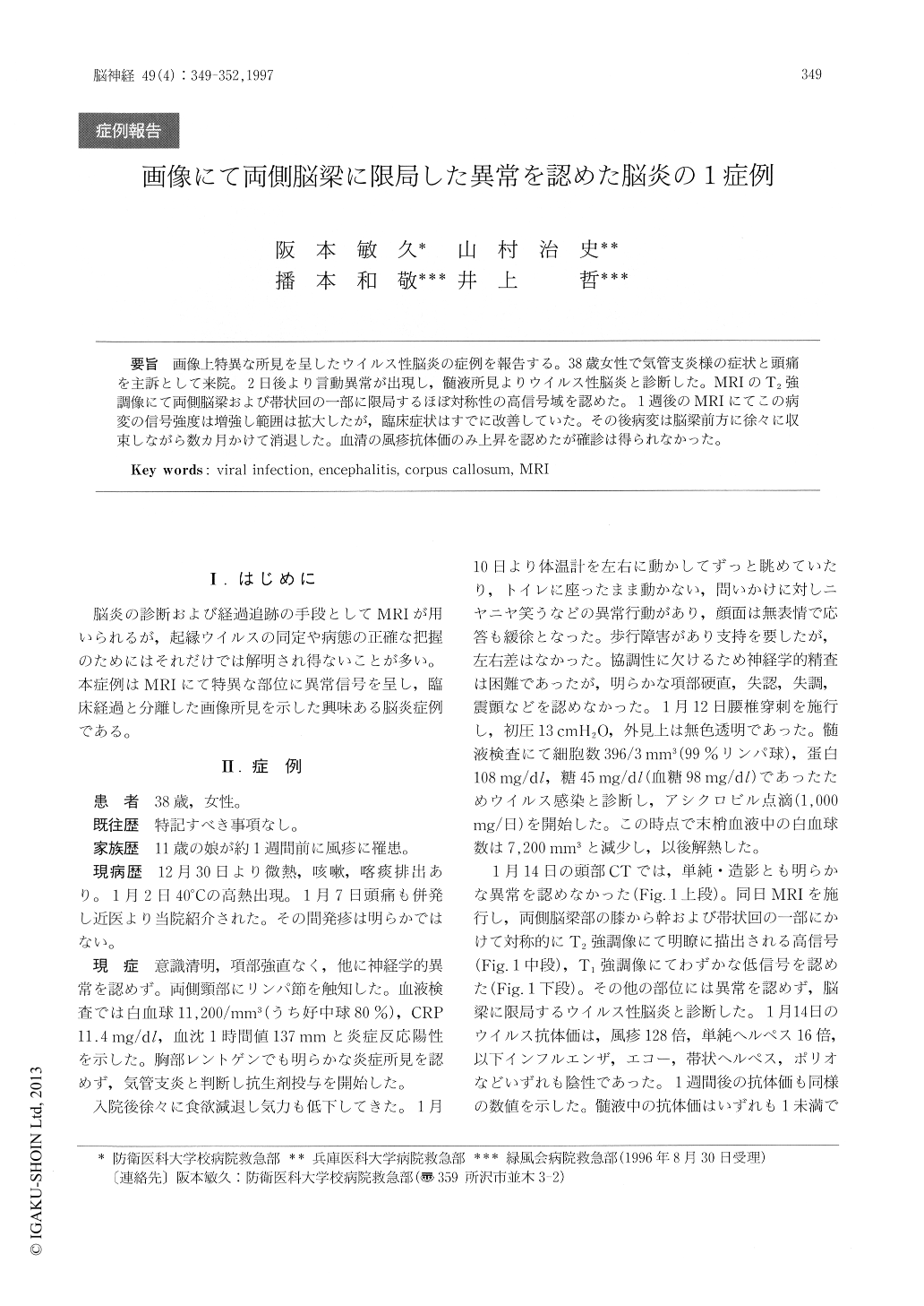
画像上特異な所見を呈したウイルス性脳炎の症例を報告する。38歳女性で気管支炎様の症状と頭痛を主訴として来院。2日後より言動異常が出現し,髄液所見よりウイルス性脳炎と診断した。MRIのT2強調像にて両側脳梁および帯状回の一部に限局するほぼ対称性の高信号域を認めた。1週後のMRIにてこの病変の信号強度は増強し範囲は拡大したが,臨床症状はすでに改善していた。その後病変は脳梁前方に徐々に収束しながら数カ月かけて消退した。血清の風疹抗体価のみ上昇を認めたが確診は得られなかった。
A case of viral encephalitis is described. A 38 year - old female was admitted because of high fever accompanied by cough and headaches. Two days after admission the patient began to exhibit abnormal behavior. Cerebrospinal fluid examination revealed pleocytosis. T2-weighted MRI revealed a high-intensity area located in the corpus callosum bilaterally that gradually increased in intensity within several days the patient's behavior returned to normal. The high-intensity area on MRI persist-ed for several months but diminished in intensity. Rubella may have been the etiology of the encepha-litis.

Copyright © 1997, Igaku-Shoin Ltd. All rights reserved.


