Japanese
English
- 有料閲覧
- Abstract 文献概要
- 1ページ目 Look Inside
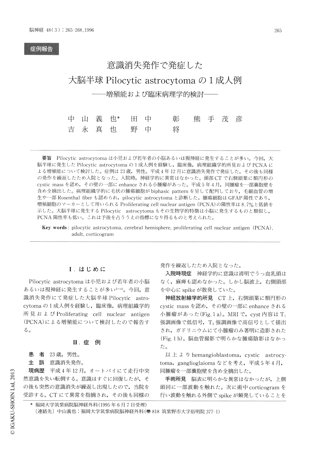
Pilocytic astrocytomaは小児および若年者の小脳あるいは視神経に発生することが多い。今回,大脳半球に発生したPilocytic astrocytomaの1成人例を経験し,臨床像,病理組織学的所見およびPCNAによる増殖能について検討した。症例は23歳,男性。平成4年12月に意識消失発作で発症した。その後も同様の発作を繰返したため入院となった。入院時,神経学的に異常はなかった。頭部CTで右側頭葉に類円形のcystic massを認め,その壁の一部にenhanceされる小腫瘤があった。平成5年4月,同腫瘤を一部嚢胞壁を含め全摘出した。病理組織学的に毛状の腫瘍細胞がbiphasic patternを呈して配列しており,毛細血管の増生や一部Rosenthal fiberも認められ,pilocytic astrocytomaと診断した。腫瘍細胞はGFAP陽性であり,増殖細胞のマーカーとして用いられるProliferating cell nuclear antigen(PCNA)の陽性率は8.7%と低値を示した。大脳半球に発生するPilocytic astrocytomaもその生物学的特徴は小脳に発生するものと類似し,PCNA陽性率も低い。これは予後を占ううえの指標になり得るものと考えられた。
The clinical and the pathological features of a surgical case of adult pilocytic astrocytoma in the right temporal lobe are described. The growth kinetics of the tumor cells were investigated by immunohistochemical staining of Proliferating cell nuclear antigen (PCNA). The patient, a 23-year-old man, was admitted to our hospitical with a history of loss of consciousness. A CT scan showed a cystic lesion with enhanced mural nodule in the right temporal lobe. Total resection of the mural nodule including the surrounding cyst wall was performed.

Copyright © 1996, Igaku-Shoin Ltd. All rights reserved.


