Japanese
English
- 有料閲覧
- Abstract 文献概要
- 1ページ目 Look Inside
- 参考文献 Reference
Ⅰ.はじめに
Pilomyxoid astrocytoma(PMA)はbiphasic patternをとるpilocytic astrocytoma(PA)とは異なり,背景はmonophasicなmyxoid patternをとる腫瘍として1999年にTihanらにより提唱された19).2007年のWHO脳腫瘍分類では,PMAはPAと比べて乳幼児に発生し予後も悪いことからGrade Ⅱとして分類されている17).しかしながらPMAは再発時にPAにmatureしたという報告が散見され1,12),2010年にJohnsonらは両者は一連のspectrum上にあるとして“intermediate pilomyxoid tumors(IPTs)”の概念を提唱している12).
一方,PAではBRAF遺伝子異常(KIAA1549-BRAF fusion)が約70%にみられるが3,4,7,8,11,15),PMAでの報告は少ない.今回われわれはPMAおよびIPT,すなわちpilomyxoid-spectrum astrocytoma(PMSA)と考えられた2例を経験したので,その臨床病理像をBRAF遺伝子異常の検討も加え報告する.
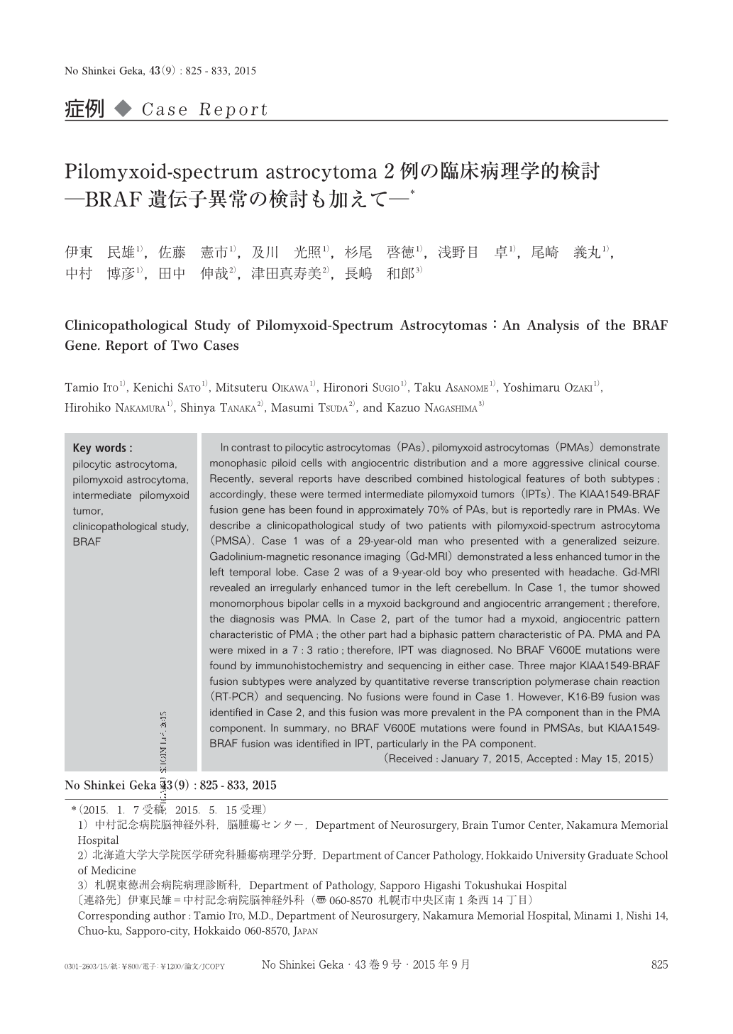
In contrast to pilocytic astrocytomas(PAs), pilomyxoid astrocytomas(PMAs)demonstrate monophasic piloid cells with angiocentric distribution and a more aggressive clinical course. Recently, several reports have described combined histological features of both subtypes;accordingly, these were termed intermediate pilomyxoid tumors(IPTs). The KIAA1549-BRAF fusion gene has been found in approximately 70% of PAs, but is reportedly rare in PMAs. We describe a clinicopathological study of two patients with pilomyxoid-spectrum astrocytoma(PMSA). Case 1 was of a 29-year-old man who presented with a generalized seizure. Gadolinium-magnetic resonance imaging(Gd-MRI)demonstrated a less enhanced tumor in the left temporal lobe. Case 2 was of a 9-year-old boy who presented with headache. Gd-MRI revealed an irregularly enhanced tumor in the left cerebellum. In Case 1, the tumor showed monomorphous bipolar cells in a myxoid background and angiocentric arrangement;therefore, the diagnosis was PMA. In Case 2, part of the tumor had a myxoid, angiocentric pattern characteristic of PMA;the other part had a biphasic pattern characteristic of PA. PMA and PA were mixed in a 7:3 ratio;therefore, IPT was diagnosed. No BRAF V600E mutations were found by immunohistochemistry and sequencing in either case. Three major KIAA1549-BRAF fusion subtypes were analyzed by quantitative reverse transcription polymerase chain reaction(RT-PCR)and sequencing. No fusions were found in Case 1. However, K16-B9 fusion was identified in Case 2, and this fusion was more prevalent in the PA component than in the PMA component. In summary, no BRAF V600E mutations were found in PMSAs, but KIAA1549-BRAF fusion was identified in IPT, particularly in the PA component.

Copyright © 2015, Igaku-Shoin Ltd. All rights reserved.


