Japanese
English
- 有料閲覧
- Abstract 文献概要
- 1ページ目 Look Inside
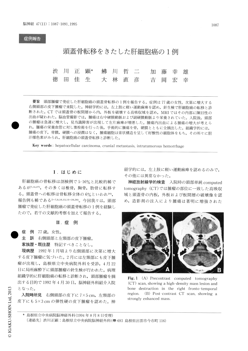
頭部腫瘤で発症した肝細胞癌の頭蓋骨転移の1例を報告する。症例は77歳の女性。次第に増大する右側頭部の皮下腫瘤で来院した。神経学的には,左上肢に軽い運動麻痺を認め,針生検で肝細胞癌の転移と診断された。CTでは頭蓋骨の板間層から内,外板を破壊する高吸収域を認め,MRIではその内部に陳旧性の出血が疑われた。脳血管撮影では,腫瘍は右中硬膜動脈および副硬膜動脈より栄養されていた。入院後,頭部の腫瘤は急速に増大し,見当識障害が出現して左片麻痺が増悪した。腫瘍内出血による腫瘍の増大が考えられ,腫瘍の栄養血管に対し塞栓術を行った後,手術的に腫瘍を骨,硬膜とともに全摘出した。組織学的には,腫瘍の皮下,骨膜,硬膜への浸潤はなく,腫瘍細胞は索状構造を呈して好酸性の細胞体をもち,その所々に胆汁様色素がみられ,肝細胞癌の頭蓋骨転移と診断した。
A case of cranial metastasis of hepatocellular carcinoma is reported.
A 77-year-old woman with an elastic hard tumor in the right temporal region was referred to our department on April 30, 1992. On admission, the patient had slight weakness of the left upper limb. Plain skull X-ray and computed tomography (CT) showed bone destruction in the right temporal region. Magnetic resonance images (MRI) showed that the tumor was hypo-intense with T1-sequences and hyper-intense with T2 sequences, and included hyper-intense spots on both T1- and T2-images.

Copyright © 1995, Igaku-Shoin Ltd. All rights reserved.


