Japanese
English
- 有料閲覧
- Abstract 文献概要
- 1ページ目 Look Inside
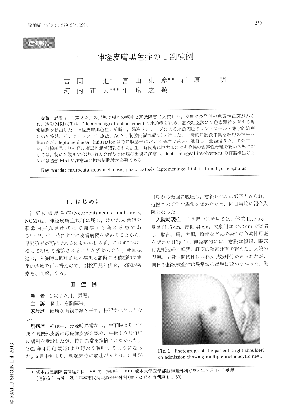
患者は,1歳2ヵ月の男児で頻回の嘔吐と意識障害で入院した。皮膚に多発性の色素性母斑がみられ,造影MRI(CT)にてleptomenigeal enhancementと水頭症を認め,髄液細胞診にて色素顆粒を有する異常細胞を検出した。神経皮膚黒色症と診断し,髄液ドレナージによる頭蓋内圧のコントロールと集学的治療(DAV療法,インターフェロン療法,ACNU髄腔内灌流療法)を行った。一時的に髄液中異常細胞の消失を認めたが,leptomeningeal infiltrationは特に脳底部において高度で急速に進行し,全経過5カ月で死亡した。剖検所見より神経皮膚黒色症が確認された。生下時皮膚に巨大または多発性の色素性母斑を認める児に対しては,特に2歳まではけいれん発作や水頭症の出現に注意し,leptomenigeal involvementの有無検出のためには造影MRIや注意深い髄液細胞診が必要である。
Neurocutaneous melanosis is a rare congenital phacomatosis characterized by the presence of large or multiple congenital melanocytic nevi and benign or malignant pigmented cell tumors of the lep-tomeninges.
A 14-month-old boy was admitted with a recent history of vomiting and drowsiness. He was found to have multiple congenital melanocytic nevi. Gd-enhanced MRI showed ventriculomegaly and lep-tomeningeal enhancement in the ambient cistern. CSF cytology revealed abnormal cells with pigment-ed granules. A diagnosis of hydrocephalus with malignant neurocutaneous melanosis was made.

Copyright © 1994, Igaku-Shoin Ltd. All rights reserved.


