Japanese
English
- 有料閲覧
- Abstract 文献概要
- 1ページ目 Look Inside
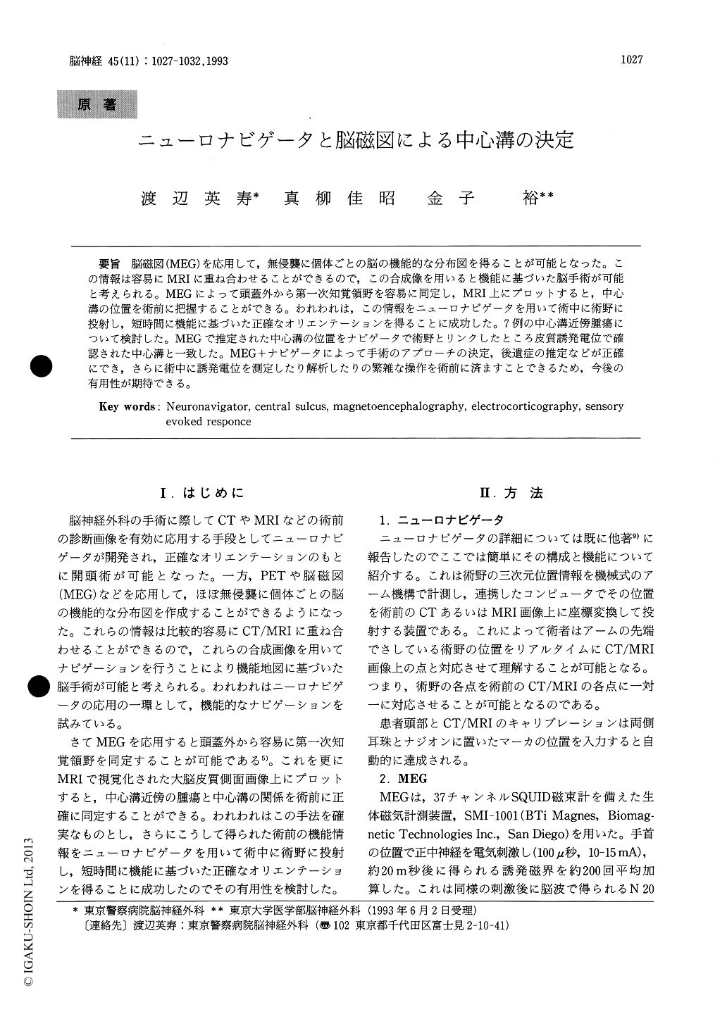
脳磁図(MEG)を応用して,無侵襲に個体ごとの脳の機能的な分布図を得ることが可能となった。この情報は容易にMRIに重ね合わせることができるので,この合成像を用いると機能に基づいた脳手術が可能と考えられる。MEGによって頭蓋外から第一次知覚領野を容易に同定し,MRI上にプロットすると,中心溝の位置を術前に把握することができる。われわれは,この情報をニューロナビゲータを用いて術中に術野に投射し,短時間に機能に基づいた正確なオリエンテーションを得ることに成功した。7例の中心溝近傍腫瘍にっいて検討した。MEGで推定された中心溝の位置をナビゲータで術野とリンクしたところ皮質誘発電位で確認された中心溝と一致した。MEG+ナビゲータによって手術のアプローチの決定,後遺症の推定などが正確にでき,さらに術中に誘発電位を測定したり解析したりの繁雑な操作を術前に済ますことできるため,今後の有用性が期待できる。
The brain-generated currents that produce poten-tials measured by the electroencephalogram also produce magnetic fields which can be measured by the magnetoencephalogram (MEG), N 20 compati-ble evoked field after median nerve stimulation is known to be generated in primary sensory cortex.Using MEG with 37 channel SQUIDs, a current dipole is back traced which corresponds to the sensory cortex. When the dipole is projected onto the MRI of the same patient, the primary sensory cortex is precisely identified in the MRI images. These data were used as the key images for naviga-tor enabling a surgeon identify the central cortex in the surgical field. Seven patients with peri-central mass lesion (3 meningiomas, 1 metastatic tumors, 1 angiomas, 2 gliomas) underwent surgery under MEG-navigator method. In every case, the central sulcus and motor cortex were easily identified on the cortex and the tumor was removed as far as possible preserving the motor strip. There were no postoperative worsening of the motor paresis and no other complications were noticed. The method which combines the MEG functional mapping and navigator was considered to be a powerful tool in surgery of the pericentral mass lesions.

Copyright © 1993, Igaku-Shoin Ltd. All rights reserved.


