Japanese
English
- 有料閲覧
- Abstract 文献概要
- 1ページ目 Look Inside
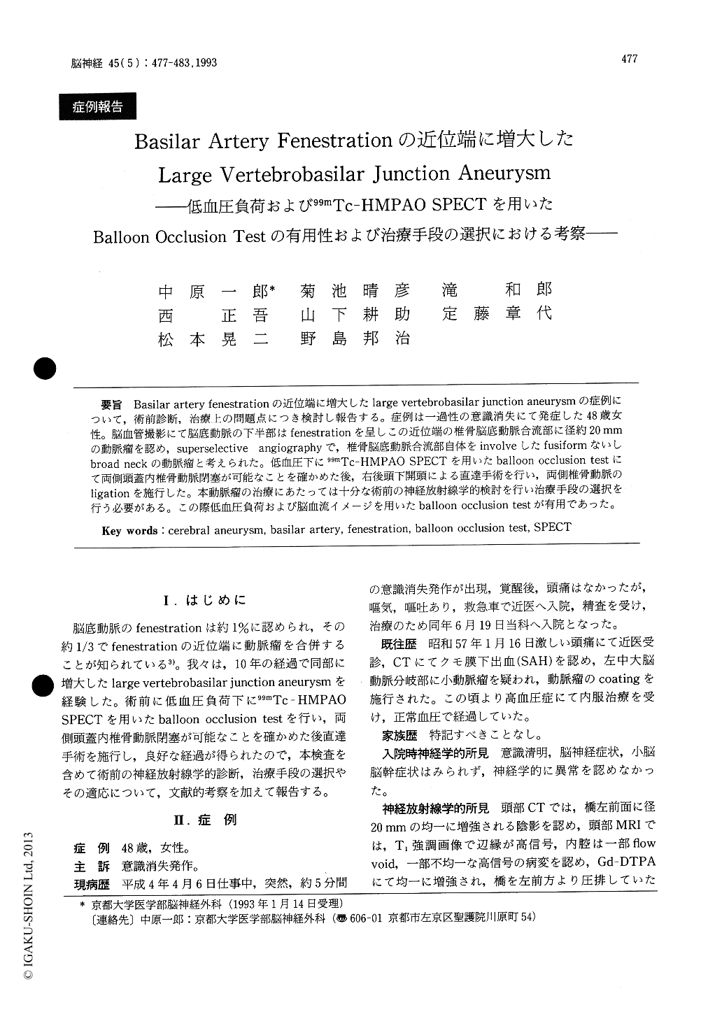
Basilar artery fenestrationの近位端に増大したlarge vertebrobasilar junction aneurysmの症例について,術前診断,治療上の問題点につき検討し報告する。症例は一過性の意識消失にて発症した48歳女性。脳血管撮影にて脳底動脈の下半部はfenestrationを呈しこの近位端の椎骨脳底動脈合流部に径約20mmの動脈瘤を認め,superselective angiographyで,椎骨脳底動脈合流部自体をinvolveしたfusiformないしbroad neckの動脈瘤と考えられた。低血圧下に99mTc-HMPAO SPECTを用いたballoon occlusion testにて両側頭蓋内椎骨動脈閉塞が可能なことを確かめた後,右後頭下開頭による直達手術を行い,両側椎骨動脈のligationを施行した。本動脈瘤の治療にあたっては十分な術前の神経放射線学的検討を行い治療手段の選択を行う必要がある。この際低血圧負荷および脳血流イメージを用いたballoon occlusion testが有用であった。
A 48-year-old lady suffered a transient loss of consciousness. CT and MRI revealed a large vascu-lar lesion compressing the left lower pons. Angio-graphy revealed a large aneurysm at vertebro-basilar junction, dome of which projected anteri-orly and left to midline. Her previous vertebral angiogram taken 10 years ago when she suffered a subarachnoid hemorrhage from the left MCA aneu-rysm, had showed a fenestration of lower basilar artery without apparent aneurysm. Bilateral super-selective vertebral angiograms revealed that the aneurysm arose at the proximal end of the fenestra-tion, and vertebrobasilar junction was incorporated into the aneurysm indicating broad neck aneurysm. The left posterior communicating artery was well developed. Balloon test occlusion (BTO) of bila-teral vertebral artery was performed under nor-motension and induced hypotension. 99mHM - PAO SPECT was used to examine cerebral blood flow (CBF) during hypotensive BTO. The patient tolera-ted the test and CBF imaging showed insignificant sight decrease in bilateral cerebellar hemispheres. Exploration of the aneurysm was carried out by the right far lateral suboccipital approach . Bilateral vertebral arteries and the right segment of the basilar artery fenestration were identified. Neck clipping of the aneurysm with reconstruction of the parent vessels were tried with fenestrate clip. How-ever, narrow operative field and large dome of theaneurysm made it hard to identify the left segment of the fenestration. Neck clipping was given up and clipping of bilateral vertebral arteries were perfor-med distal to posterior inferior cerebellar artery with three body clippings. The patient showed moderate postoperative left lower nerve palsy, which was gradually improved in several weeks. Follow-up angiography revealed no opacification ofthe aneurysm. BTO with hypotensive challenge and CBF imaging appeared extremely useful in examin-ing possible neurological complications after perma-nent parent artery occlusion. Considerations in neu-roradiological diagnosis and therapeutic strategies for aneurysms of this type were also described.

Copyright © 1993, Igaku-Shoin Ltd. All rights reserved.


