Japanese
English
- 有料閲覧
- Abstract 文献概要
- 1ページ目 Look Inside
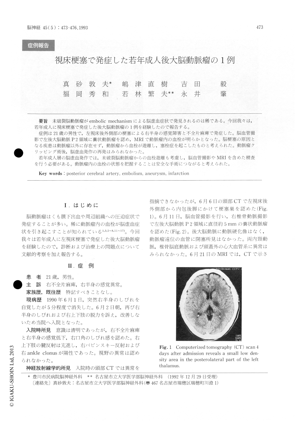
未破裂脳動脈瘤がembolic mechanismによる脳虚血症状で発見されるのは稀である。今回我々は,若年成入に視床梗塞で発症した後大脳動脈瘤の1例を経験したので報告する。
症例は21歳の男性で,左視床後外側部の梗塞による右半身の感覚障害と不全片麻痺で発症した。脳血管撮影で左後大脳動脈P2領域に嚢状動脈瘤を認め,MRIで動脈瘤内の血栓が明らかとなった。脳梗塞の原因となる疾患は動脈瘤以外に存在せず,動脈瘤から血栓が遊離し,塞栓症を起こしたものと考えられた。動脈瘤クリッピング術後,脳虚血発作の再発はみられなかった。
若年成人層の脳虚血発作では,未破裂脳動脈瘤からの血栓遊離も考慮し,脳血管撮影やMRIを含めた精査を行う必要がある。動脈瘤内の血栓の状態を把握することは安全な手術につながると考えられた。
A 21-year-old man presented with sudden weak-ness and dysesthesia of his right limbs. Computer-ized tomography (CT) scan showed a low density area in the posterolateral part of the left thalamus. Right vertebral angiography revealed a small aneu-rysm at the P2 segment of the posterior cerebral artery. Magnetic resonance imaging (MRI) demon-strated intraaneurysmal clot and signal void in the residual lumen. There were no other lesion and no predisposing risk factors that produced cerebral ischemia. It was thougt that the aneurysm was the source of emboli resulting in thalamic infarction. The patient underwent a left subtemporal crani-otomy, and the aneurysm was clipped. Following surgery, there has been no recurrence of ischemic attacks. The diagnosis and the therapy were discussed, with reference to the literature.

Copyright © 1993, Igaku-Shoin Ltd. All rights reserved.


