Japanese
English
- 有料閲覧
- Abstract 文献概要
- 1ページ目 Look Inside
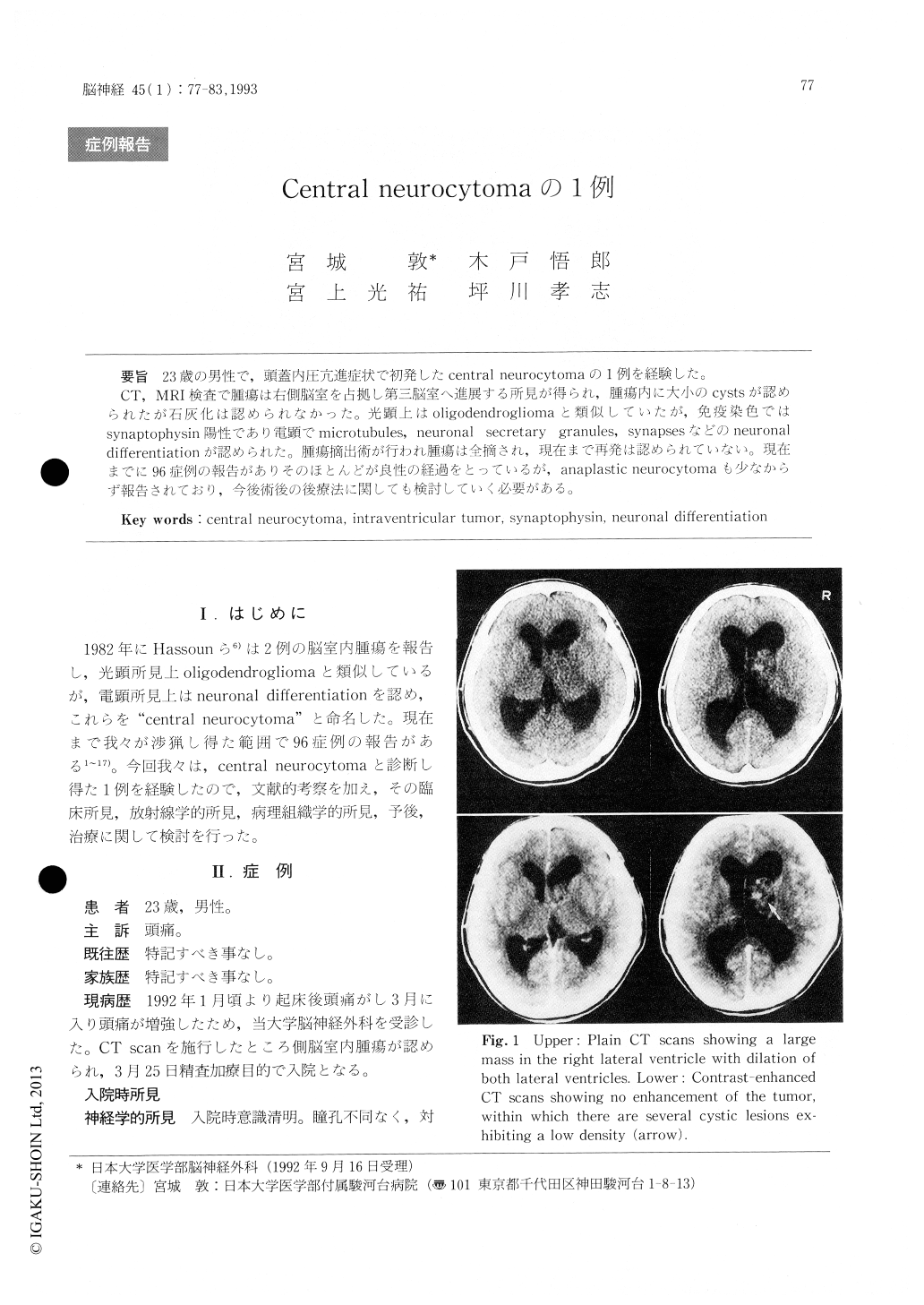
23歳の男性で,頭蓋内圧亢進症状で初発したcentral neurocytomaの1例を経験した。
CT,MRI検査で腫瘍は右側脳室を占拠し第三脳室へ進展する所見が得られ,腫瘍内に大小のcystsが認められたが石灰化は認められなかった。光顕上はoligodendrogliomaと類似していたが,免疫染色ではsynaptophysin陽性であり電顕でmicrotubules, neuronal secretary granules, synapsesなどのneuronaldifferentiationが認められた。腫瘍摘出術が行われ腫瘍は全摘され,現在まで再発は認められていない。現在までに96症例の報告がありそのほとんどが良性の経過をとっているが,anaplastic neurocytomaも少なからず報告されており,今後術後の後療法に関しても検討していく必要がある。
The authors present a case of central neuro-cytoma in a 23-year-old male with increased intra-cranial pressure syndrome. Computed tomographic (CT) scans and magnetic resonance images showed a large tumor mass with no evidence of calcification in the right lateral ventricle extending towards the third ventricle. A right transcortical - transvent-ricular approach was performed and the tumor was totally removed. The postoperative course was uneventful and no further treatment was administe-red. CT shows no evidence of tumor recurrence after the six months from his surgery. Light microscopic findings suggested a diagnosis of oligodendrogli-oma. However, ultrastructural examinations demon-strated many dense-core or clear vesicles, micro-tubules and synaptic like structures within the abundant cytoplasmic processes of the tumor cellswhich suggested neuronal differentiation. Immuno-histochemical examinations showed the tumor cells to be positive for neuron-specific enolase, sporadi-cally positive for synaptophysin, and negative for glial fibrillary acidic protein. The final histological diagnosis was central neurocytoma. Central neuro-cytoma was first described by Hassoun et al, in1982. Since then, 96 cases have been reported in the literatures. Their clinicopathological features, neuro-radiological findings and prognosis are discussed.

Copyright © 1993, Igaku-Shoin Ltd. All rights reserved.


