Japanese
English
- 有料閲覧
- Abstract 文献概要
- 1ページ目 Look Inside
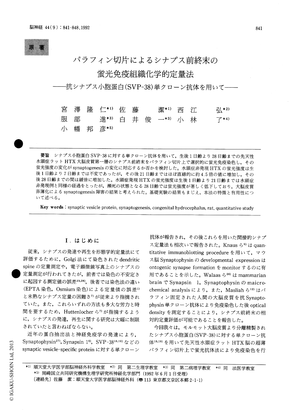
シナプス小胞蛋白SVP−38に対する単クローン抗体を用いて,生後1日齢より28日齢までの先天性水頭症ラットHTX大脳皮質第一層のシナプス前終末をパラフィン切片上で選択的に蛍光免疫染色し,その蛍光強度の変化がsynaptogenesisの変化に対応するか否かを検討した。水頭症非発現HTXの蛍光強度は生後1日齢より7日齢までは不変であったが,その後21日齢まではほぼ直線的に約4.5倍の値に増加し,その後28日齢までの間は緩徐に増加した。水頭症発現HTXの蛍光強度は生後1日齢より21日齢までは水頭症非発現例と同様の経過をとったが,瀕死の状態となる28日齢では蛍光強度が著しく低下しており,大脳皮質菲薄化によるsynaptogenesis障害の結果と考えられた。基礎実験の結果もまじえ,本法の特徴と有用性について述べる。
We developed a method for investigating impair-ment of synaptogenesis quantitatively involving measurement of the fluorescenece intensity emitted by immunohistochemically stained paraffin sections of rat brain using a monoclonal antibody (Mab 171B5) against a synaptic vesicle protein (SVP-38). We applied this method to congenitally hydroce-phalic and non-hydrocephalic brains of HTX-rats, and compared the postnatal changes in the fluores-cence intensity in the molecular layer of the cere-bral cortex. In non-hydrocephalic HTX-rats, the fluorescence intensity remained nearly unchanged from the 1st to 7th postnatal day and then increased at an almost linear rate until the 21st postnatal day, when it reached 4.5 times the value on the 7th postnatal day. The increase thereafter was gradual until the 28th postnatal day. In hydrocephalic HTX -rats, the fluorescence intensity showed a marked reduction on the 28th postnatal day (p<0.01). This finding indicated impairment of synaptogenesis. We believe that this method provides useful information for evaluating the impairment of synaptogensis in various pathological conditions in mammarian brains. The basic aspects of this method which support its validity are also discussed.

Copyright © 1992, Igaku-Shoin Ltd. All rights reserved.


