Japanese
English
- 有料閲覧
- Abstract 文献概要
- 1ページ目 Look Inside
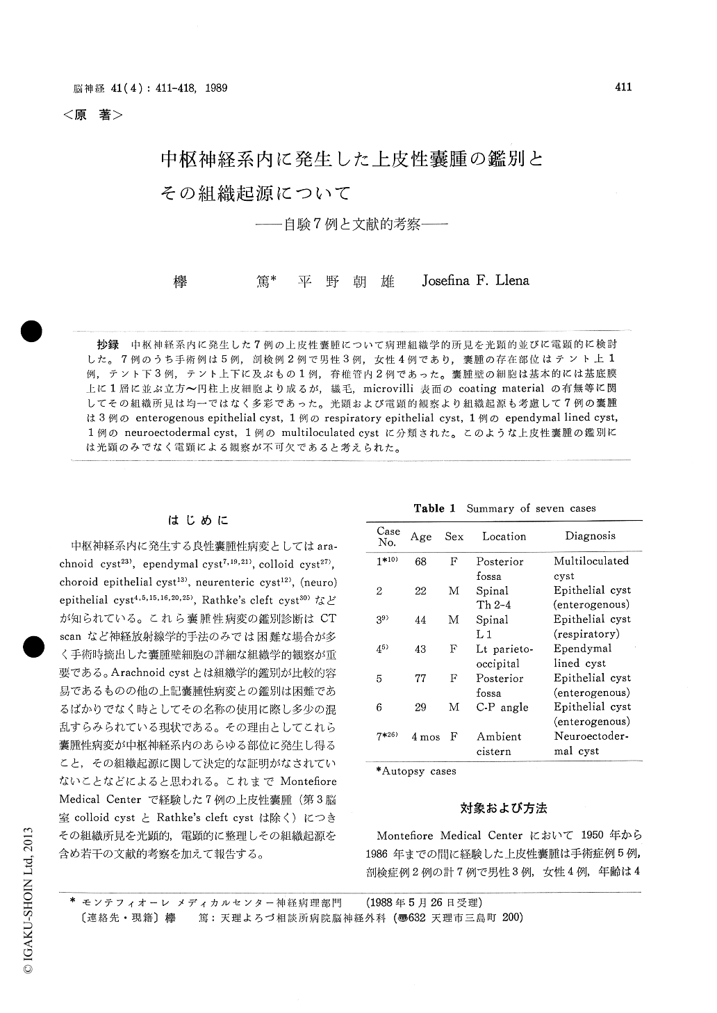
抄録 中枢神経系内に発生した7例の上皮性嚢腫について病理組織学的所見を光顕的並びに電顕的に検討した。7例のうち手術例は5例,剖検例2例で男性3例,女性4例であり,嚢腫の存在部位はテント上1例,テント下3例,テント上下に及ぶもの1例,脊椎管内2例であった。嚢腫壁の細胞は基本的には基底膜上に1層に並ぶ立方〜円柱上皮細胞より成るが,繊毛,microvilli表面のcoating materialの有無等に関してその組織所見は均一ではなく多彩であった。光顕および電顕的観察より組織起源も考慮して7例の嚢腫は3例のenterogenous epithelial cyst,1例のrespiratory epithelial cyst,1例のependymal lined cyst,1例のneuroectodermal cyst,1例のmultiloculated cystに分類された。このような上皮性?腫の鑑別には光顕のみでなく電顕による観察が不可欠であると考えられた。
Seven cases of epithelial cysts are presented with special reference to histological findings. Dif-ferential diagnosis and origin of the cysts are also discussed.
Two are autopsy cases and 5 are surgical cases. Median age of the patients is 41 years. Three cysts are in the posterior fossa, 1 in the supratentorial region, 1 in both infra- and supra-tentorial regions and 2 in the spinal canal. On light microscopy, the type of cell lining the cyst wall and the presenece of cilia and PAS-positive cells are studied. All cyst walls were lined by a single layer of cuboidal to columnar epithelium. Cilia was seen in 1 and PAS-positive cells were found in 5 out of 7 cases.
On electron microscopy of the 4 cases available for study, continuous basement membrane and microvilli were observed in all cases. Coating material covering microvilli was noted in 2 cases.
According to these histological findings, these cysts are classified as follows : 1 multiloculated cyst, 1 (respiratory) epithelial cyst, 3 (enteroge-nous) epithelial cysts, 1 ependymal lined cyst and 1 neuroectodermal cyst.
Various non-neoplastic cystic lesions are found in the central nervous syetem, such as arachnoid cyst, ependymal cyst, colloid cyst, choroid epithe-lial cyst, neurenteric cyst, and Rathke's cleft cyst. Although histological difference between arach-noid cyst and other epithelium-lined cysts is re-latively clear, the precise discrimination between other cystic lesions is difficult and controversial. Some authors have considered these cysts as a neuroectodermal origin because of their histologi-cal similality with choroid plexus or ependyma.
On the other hand, others have proposed an endo-dermal origin based on histological close resem-blance to respiratory or gastrointestinal tracts epi-thelial cells on electron microscopic features. Ac-cording to our study, we propose that names of epi-thelial cyst should be used when cyst wall is com-posed of a single layer of epithelial cells with a continuous basement membrane and microvilli covered with an electon dense materials. Ependy-mal cyst is basically distinguished from our epi-thelial cyst on the absence of basement membrane and PAS-positive cell, however, some variable histological patterns may exist. The definite di-agnosis should be made based on both anatomical location and histological findings with electron microscopic study.
This report demonstrates the limitations of light microscopy in distinguishing the precise nature of the cysts lining epithelium, and the capability of the electron microscope in defining the fine struc-ture for certain epithelium.
Copyright © 1989, Igaku-Shoin Ltd. All rights reserved.