Japanese
English
- 有料閲覧
- Abstract 文献概要
- 1ページ目 Look Inside
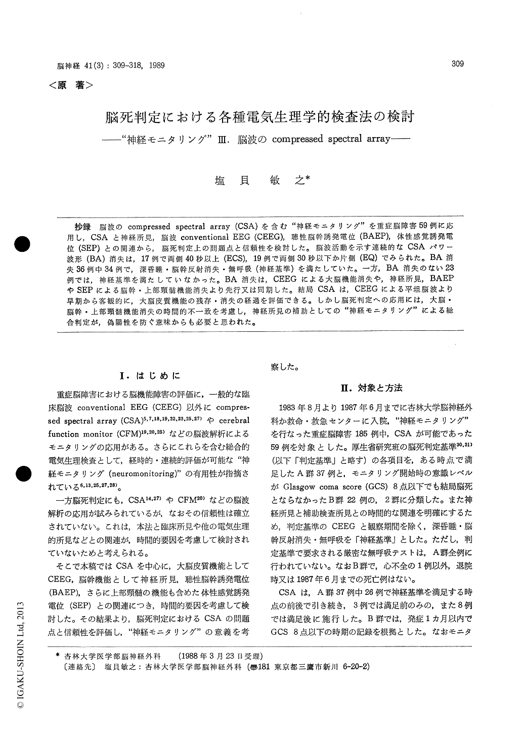
抄録 脳波のcompressed spectral array (CSA)を含む"神経モニタリング"を重症脳障害59例に応用し,CSAと神経所見,脳波conventlonal EEG (CEEG),聴性脳幹誘発電位(BAEP),体性感覚誘発電位(SEP)との関連から,脳死判定上の問題点と信頼性を検討した。脳波活動を示す連続的なCSAパワー波形(BA)消失は,17例で両側40秒以上(ECS),19例で両側30秒以下か片側(EQ)でみられた。BA消失36例中34例で,深昏睡・脳幹反射消失・無呼吸(神経基準)を満たしていた。一方,BA消失のない23例では,神経基準を満たしていなかった。BA消失は,CEEGによる大脳機能消失や,神経所見,BAEPやSEPによる脳幹・上部頸髄機能消失より先行又は同期した。結局CSAは,CEEGによる平坦脳波より早期から客観的に,大脳皮質機能の残存・消失の経過を評価できる。しかし脳死判定への応用には,大脳・脳幹・上部頸髄機能消失の時間的不一致を考慮し,神経所見の補助としての"神経モニタリング"による総合判定が,偽陽性を防ぐ意味からも必要と思われた。
Electrophysiological neuromonitoring of com-pressed spectral array (CSA) EEG, brainstem audi-tory evoked potentials (BAEP) and short-latency somatosensory evoked potentials (SEP) can provide precise and immediate information concerning functional integrity of severe brain damage.
We applied this neuromonitoring system in 185 cases of severe brain damage in order to evaluate its reliability in the diagnosis of brain death. This report considers the relationships between CSA and neurological, conventional EEG (CEEG), BAEP and SEP findings, and the significance of CSA in the assessment of brain death.
CSA monitoring of 59 patients (37 brain-dead ingroup A and 22 non brain-dead in group B) was performed and analysed.
Brain damage was caused by cerebrovascular insult in 31 cases, head injury in 25, meningitis in 2, dan anoxia in 1. Mean patient age was 49 (ages 5-84). There were no significant differences in causes and age distribution between the two groups.
CSA monitoring, using 5 or 7 μV/mm, of two channels (Cz-A 1 and Cz-A 2) of EEG activity was performed. Power spectral analysis (0-16 Hz) was carried out at 10 to 120 second epochs.
Automatic BAEP (54 patients) and/or SEP (33 patients) monitoring was performed simultaneously 10 to 30 minutes.
CSA was classified into three patterns (Fig. 1) : 1) "Electrocerebral silence ; ECS" revealed bilate-ral absence of the CSA power spectra for periods longer than 40 seconds.
2) "Biological activity ; BA" showed continuous peaks of activity.
3) "Equivocal ; EQ" pattern showed intermittent peaks of activity, and unilateral loss of power spectra or bilateral loss within 30 seconds.
CSA findings indicated the loss of CSA "BA" pattern (LCBP) in 36 group A patients (17 "ECS" and 19 "EQ").
All but two revealing LCBP had satisfied the neurological criteria of brain death (deep coma, absent brainstem reflexes and apnea) during CSA monitoring. "BA" was always present in 22 group B and one group A patients had not fulfilled neuro-logical criteria of brain death.
The CEEG results of all group A patients demon-strating "ECS" of CSA were of the classification Hockaday grade V a or V b. Four patients clas-sified as grade N b demonstrated "EQ" (Table 1 and Fig. 2 A).
An examination of CSA and neurological brain-stem function of 28 group A patients revealed LCBP preceded the loss of brainstem reflexes and apnea in 15 patients or was coincided with them in 13 (Table 2).
CSA and BAEP findings of 33 group A patients (Table 3) indicated :
( 1 ) LCBP preceding the loss of BAEP wave 11 - V in 12 patients (Fig. 3);
( 2 ) LCBP associated with this loss in 12 patients (Fig. 2B);
( 3 ) LCBP coincided with the loss of all BAEP in 9 patients.
CSA and SEP findings in 20 group A patients indicated (Table 4):
( 1 ) LCBP preceding the loss of scalp P 13-14 with earlobe reference in 3 patients (Fig. 3);
( 2 ) LCBP associated with loss of scalp P 13-14 with earlobe reference, or scalp P 15 with Fz refer-ence, in remaining 3 patients.
Cerivical N 14, however, was preserved after LCBP in 5 patients.
Meanwhile, a patient in group B who had lost all BAEP waves and cervical N 13, N 14 and scalp P 15 of SEP with Fz reference demonstrated bio-logical activity on CSA and CEEG (Fig. 4).
In other words, CSA is more objective and quicker in indicating the persistence or disappear-ance of cerebral function than CEEG.
Taking into consideration the temporal relation-ships between functional loss in the cerebrum, brainstem, and upper spinal cord, it is suggested that neuromonitoring is required for elimination of false-positive and false-negative results based on only one electrophysiological test and the ulti-mate diagnosis of brain death.

Copyright © 1989, Igaku-Shoin Ltd. All rights reserved.


