Japanese
English
- 有料閲覧
- Abstract 文献概要
- 1ページ目 Look Inside
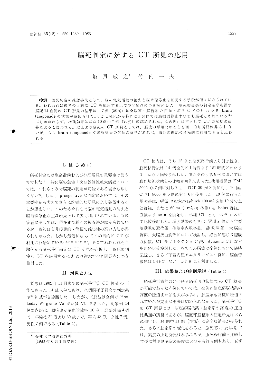
抄録 脳死判定の確認手段として,脳の電気活動の消失と脳循環停止を証明する手段が種々試みられている。われわれは後者の目的にCTを応用する上での問題点につき検討した。脳死委員会の判定基準を満す脳死14症例のCT所見の結果は,7例(50%)に全脳室・脳槽系の圧迫・消失などのいわゆるbraintamponadeの状態が認められた。しかし従来から特に欧州諸国では脳循環停止すなわち脳死とされている28)にもかかわらず,増強効果はなお10例中7例(70%)に認められた。この理由は主としてCTの感度の改善によると思われる。以上より脳死のCT所見としては,脳波の平坦化のごとき画一的な所見は得られないが,もしbrain tamponadeや増強効果の欠如の所見があれば,脳死の確認に積極的に利用できると思われる。
The absence of cerebral circulation and electro-cerebral silence have served as an accurate index of irreversible brain death.
It is proposed that computed tomography (CT) findings be evaluated as confirmatory criteria of brain death. To this end, CT evaluation of 14 pa-tients satisfying the conventional criteria of brain death was performed.
A CT finding of severe compression or dissap-pearance of the ventricular system, or so-called "brain tamponade", was seen in 7 (50%) of the 14 patients.
Enhanced contrast CT, especially dynamic CT,usually distinctly reveals the cerebral vessels when-ever the cerebral blood flow is preserved ; con-versely, the lack of enhanced brain structures, even comparing attenuation values, indicates the absence of cerebral blood flow.
In 7 (70%) of 10 patients, however, there was enhanced contrast of vascular brain structures, especially the circle of Willis, major cerebral arteries, choroid plexuses, and venous sinuses. It is suggested that this result is due to the impro-vement of demonstrability by CT.
The usefulness of CT in the confirmation of brain death lies in visualization of the pathological changes associated with a dead brain, such as "brain tamponade", and the lack of enhanced contrast indicating the absence of cerebral blood flow. The latter point is still problematic as angiography revealed an extremely low cerebral blood flow in a few cases of "dead brain" patients.
It is recommended that cerebral blood flow in brain death be evaluated by dynamic CT scanning and correlated with other methods of cerebral blood flow determination (e. g., intravenous digital subtraction angiography).

Copyright © 1983, Igaku-Shoin Ltd. All rights reserved.


