Japanese
English
- 有料閲覧
- Abstract 文献概要
- 1ページ目 Look Inside
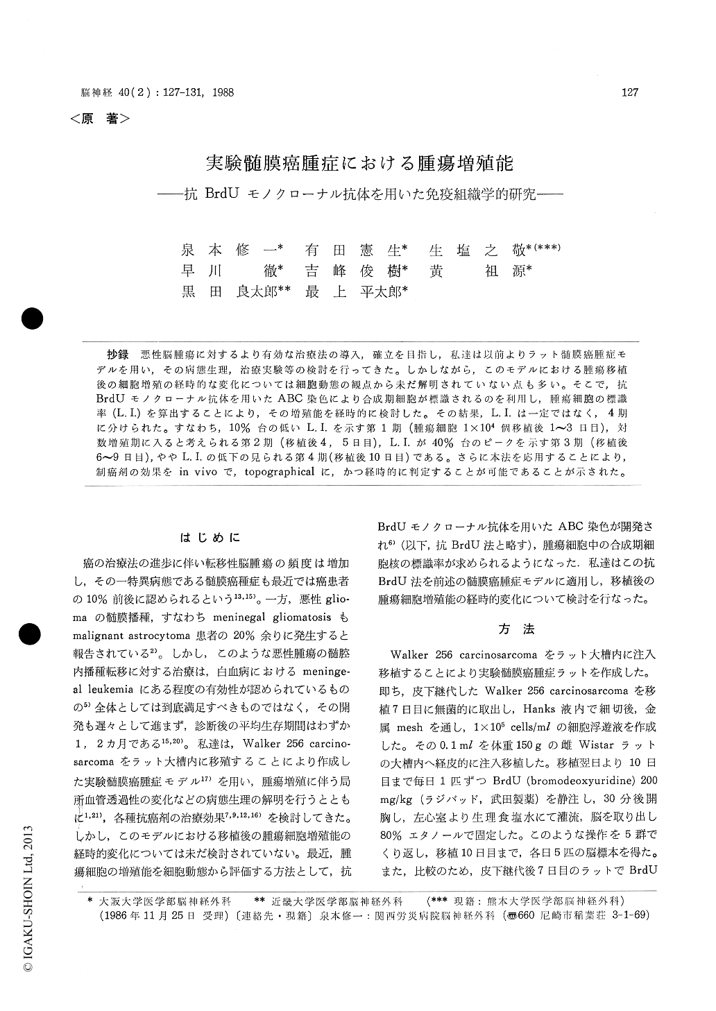
抄録 悪性脳腫瘍に対するより有効な治療法の導入,確立を目指し,私達は以前よりラット髄膜癌腫症モデルを用い,その病態生理,治療実験等の検討を行ってきた。しかしながら,このモデルにおける腫瘍移植後の細胞増殖の経時的な変化については細胞動態の観点から未だ解明されていない点も多い。そこで,抗BrdUモノクローナル抗体を用いたABC染色により合成期細胞が標識されるのを利用し,腫瘍細胞の標識率(L.I.)を算出することにより,その増殖能を経時的に検討した。その結果,L.I.は一定ではなく,4期に分けられた。すなわち,10%台の低いL.I.を示す第1期(腫瘍細胞1×10⁴個移植後1〜3日目),対数増殖期に入ると考えられる第2期(移植後4,5日目),L.I.が40%台のピークを示す第3期(移植後6〜9日目),ややL.I.の低下の見られる第4期(移植後10日目)である。さらに本法を応用することにより,制癌剤の効果をin vivoで,topographicalに,かつ経時的に判定することが可能であることが示された。
We have developed an experimental model of leptomeningeal tumor by inoculating Walker 256 carcinosarcoma cells into the cisterna magna of rats. This model was considered to be useful in studying pathophysiology and treatment of malig-nant brain tumors. In this study, the growth kine-tics of this experimental tumor was investigated by using the immunohistochemical technique with an anti-BrdU monoclonal antibody. Walker 256 carcinosarcoma was subcutaneously passaged in female Wistar rats. Seven days after subcutaneous inoculation, the tumor was aseptically removed and minced in Hank's medium by scissors to make single cell suspension of the tumor. The cell sus-pension was adjusted to 1×10⁵ cells/ml. And 0.1 ml was inoculated percutaneously into the cisterna magna of female Wistar rats weighing 150 gr. Every day after tumor inoculation, the animal (5 on each day) was sacrificed 30 minutes after in-travenous BrdU (200 mg/kg) and perfused by saline. Then, the brain was removed, fixed in ethanol and embedded in paraffin. Coronal sections of the brain 6μ in thickness were cut and stained by the indirect immunoperoxidase (ABC) method. The anti-BrdU monoclonal antibody (Becton-Dickinson) was diluted in 1 : 100. The sections were counter-stained by hematoxylin. Labelling index (L. I.) of the tumor was obtained by counting immuno-reactive cells under the microscope. L. I. of the subcutaneous tumor 7 days after inoculation was 52.4%. In the tumor 1 to 3 days after inoculation, L. I. was still low and between 11.9 and 15.1%. Four or 5 days after inoculation, the tumor cells grew in several layers in the subarachnoid space. L. I. at this stage of the tumor growth was 26. 6 to 34.8%. Six to 9 days after tumor inoculation, the massive tumor was growing in the subarach-noid space, and L. I. reached 37.9 to 47.7%. Ten days after inoculation, the tumor became necrotic in part and had a lower L. I. of 31.7%. Thus, in this experimental tumor, L. I. began to increase 4 to 5 days after tumor inoculation. Therefore, it is considered that the tumor entered the exponen-tial stage of the growth at the relatively earlytime after inoculation in this experimental tumor. Between 6 and 9 days after inoculation, L. I. reached a plateau and was almost the same as that of the subcutaneous tumor. It is possible that the immunohistochemical method using the anti-BrdU monoclonal antibody in our experimental model of the leptomeningeal tumor can be used as a topographical drug sensitivity test of various antineoplastic agents.

Copyright © 1988, Igaku-Shoin Ltd. All rights reserved.


