Japanese
English
- 有料閲覧
- Abstract 文献概要
- 1ページ目 Look Inside
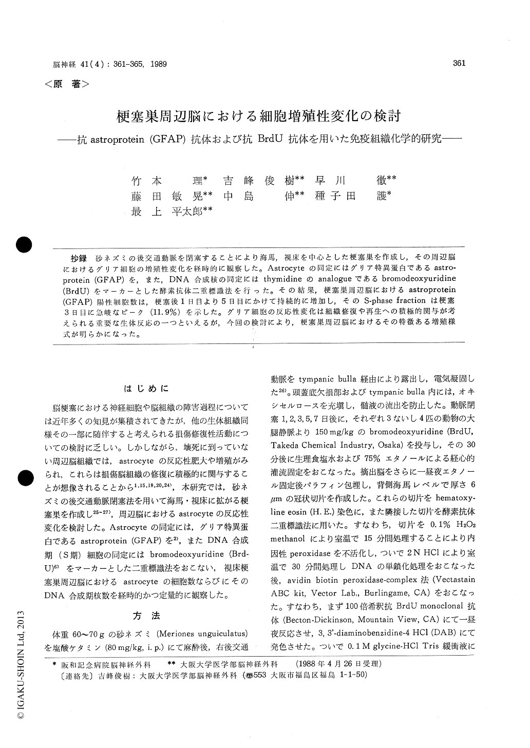
抄録 砂ネズミの後交通動脈を閉塞することにより海馬,視床を中心とした梗塞巣を作成し,その周辺脳におけるグリア細胞の増殖性変化を経時的に観察した。Astrocyteの同定にはグリア特異蛋白であるastro-protein (GFAP)を,また,DNA合成核の同定にはthymidineのanalogueであるbromodeoxyuridine(BrdU)をマーカーとした酵素抗体二重標識法を行った。その結果,梗塞巣周辺脳におけるastroprotein(GFAP)陽性細胞数は,梗塞後1日目より5日目にかけて持続的に増加し,そのS-phase fractionは梗塞3日目に急峻なピーク(11.9%)を示した。グリア細胞の反応性変化は組織修復や再生への積極的関与が考えられる重要な生体反応の一つといえるが,今回の検討により,梗塞巣周辺脳におけるその特徴ある増殖様式が明らかになった。
Although cerebral infarction is a destructive process of nerve cells and brain tissue, the nature is not exclusively disintegrating but also includes active cellular reaction which may modify the progression of tissue damage. Most prominent cellular reaction in the area surrounding infarction can be recognized as a trophic or proliferative change of glial cells. In the present study we produced a focal cerebral ischemia in Mongoliangerbils and investigated the dynamic change of astrocytes in the brain adjacent to thalamic infarction. Using immunohistochemical methods, astrocytes were identified with the antibody to astroprotein (GFAP) and the DNA synthesizing (S phase) cells were detected with the antibody to bromodeoxyuridine (BrdU). The posterior com-municating artery of a gerbil was occluded by coagulation through the trans-tympanic bulla ap-proach under general anesthesia with ketamine hydrochloride (80 mg/kg, i. p.). Thirty min after intravenous administration of BrdU (200 mg/kg), animals were sacrificed by transcardiac perfusion with 75% ethanol on days 1, 2, 3, 5 and 7 post-infarction. Ethanol-fixed, paraffin-embedded blocks were cut coronally into 6 micron-thick sections at the level of dorsal hippocampus. Double-labeled immunohistochemical technique (avidin biotin per-oxidase-complex method) was carried out with each antibody using 3, 3'-diaminobenzidine tetrahydro-chloride and 4-chloro-l-naphtol as chromogens. The population of GFAP-positive cells and their S-phase fraction (the number of BrdU-positive nuclei divided by the number of GFAP-positive cells expressed in per cent, %) were examined. The data demonstrated that the regional GFAP-positive cells increased continuously between days 1 to 5 (105. 9 to 528.8 cells/mm2) postinfarction (44. 6 cells/mm2 in normal brain). Their S-phase fraction, however, remained low till day 2 (1. 0%), showed a sharp rise at day 3 (11. 9%) and de-creased rapidly thereafter (2. 8% and 1. 1% on days 5 and 7, respectively). The present study demon-strated that the focal cerebral infarction promotes active astrocytic reaction characterized by a well-organized pattern of cellular hypertrophy and proliferation, which seems important in the pro-tective and repair process against nerve tissue damage.

Copyright © 1989, Igaku-Shoin Ltd. All rights reserved.


