Japanese
English
- 有料閲覧
- Abstract 文献概要
- 1ページ目 Look Inside
I.はじめに
実験脳腫瘍は腫瘍の発生,腫瘍に付随するさまざまな病態の解明や化学療法の検討などに欠くことのできないものである。しかしながらその基本となる病理組織学的性格については十分明らかにできない場合も少なくないといわれる15)。
astroproteinはBogochらの発見した10B蛋白の主成分で,1970年に森らによりヒト・グリオーマ組織より抽出精製されたグリア特異蛋白である1,6,7)。この蛋白性抗原に対する抗血清を用いた免疫組織的学研究により,この蛋白はヒト,ラット,マウスなどのastrogliaに特異的に存在することが証明されている8〜10)。そこで私達は抗astroprotein血清を用いた螢光抗体法を実験脳腫瘍の病理組織学的診断に応用し,この方法が有用であることを報告した17)。
Immunofluorescence method using antiserum against astroprotein (an astrocyte-specific cerebro-protein, Mori et al., 1970) demonstrates fluorescence specific to the cytoplasm of normal (fibrillary), reactive and neoplastic astroglial cells. Our previous study has demonstrated that this method is very useful for the histopathological diagnosis of ethylnitrosourea-induced rat brain tumors. This method, however, has a few disadvantages for a practical use. It is unable to keep the preparations permanently because fluorescence fades. Counter-strain is impossible. The fluorescence microscope is necessary and observation is to be done in the dark room.
In the present study we tried to apply immuno-peroxidase method for the histopathological diagnosis of rat brain tumors in order to circumvent the disadvantages of immunofluorescence method.
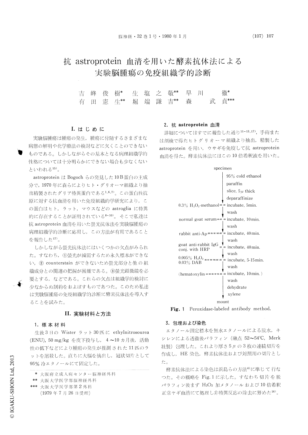
Thirty Wistar rats were subcutaneously injected with ethylnitrosourea, 50 mg/kg, on the third day of birth. They were sacrificed four to ten months later. The brain was sectioned 2 mm thick coronally, fixed in 95% cold ethanol and embedded in paraffin. Three successive coronal sections, 5μthick, were made and processed for HE stain, immunoperoxidase method and a control res-pectively. Immunoperoxidase method was carriedout at room temperature. Sections of immuno-peroxidase method were initially treated with H202- methanol and with normal goat serum in order to reduce nonspecific reactions. They were then reacted for 40 min. with rabbit anti-astroprotein serum diluted 10 folds with PBS, washed, reacted for 40 min. with peroxidase-labeled goat anti-rabbit IgG serum, washed, incubated for 5 to 15 min. in 0.05 M tris buffer, pH 7.6, containing 0.005% of H202 and 0.03% of 3, 3´-diaminobenzidine tetra-hydrochloride, washed, dehydrated, cleaned in xylol and mounted. Control sections were reacted with the normal (unimmunized) rabbit serum instead of anti-astroprotein serum.
Under the microscope, reaction products of immunoperoxidase method were observed dark brown in color. Comparative study with the control preparations revealed that the products were strictly specific to the immune reactions to astroprotein.
With the aid of immunoperoxidase method, a total number of 22 intracranial gliomas, developed in 10 rats, were diagnosed as 11 mixed gliomas of oligodendrogliomas and astrocytomas (oligoastro-cytomas), 9 oligodendrogliomas, one astrocytoma and one glioblastoma. Astrocytoma was diagnosed by the characteristic morphology of tumor cells,the cell bodies and processes of which were stained dark brown in color. Oligodendroglioma was di-agnosed by the lack of positive reactions to astro-protein. Mixed glioma of oligodendroglioma and astrocytoma (oligoastrocytoma) was diagnosed by the presence of two types of cells, oligodendroglioma cells and astrocytoma cells, in the tumor. Counter-stain with hematoxylin was especially useful to diagnose this type of tumor. A very small astro-cytoma, approximately 300μ in diameter, which was not detected in HE stain, was demonstrated by the immunoperoxidase method. Reactive astro-cytes located around oligodendroglioma were also stained by the immunoperoxidase method. Epen-dymal cells, which do not react to the anti-astroprotein serum in normal conditions, were stained dark brown when subependymal region was infiltrated by the tumor.
This study revealed the immunoperoxidase method to be superior to immunofluorescence method for several reasons. Firstly preparations by immuno-peroxidase method is kept permanently without fading of the dye. Secondly they are able to be observed under the ordinary light microscope and thirdly counterstain such as with hematoxylin is possible.
Copyright © 1980, Igaku-Shoin Ltd. All rights reserved.