Japanese
English
- 有料閲覧
- Abstract 文献概要
- 1ページ目 Look Inside
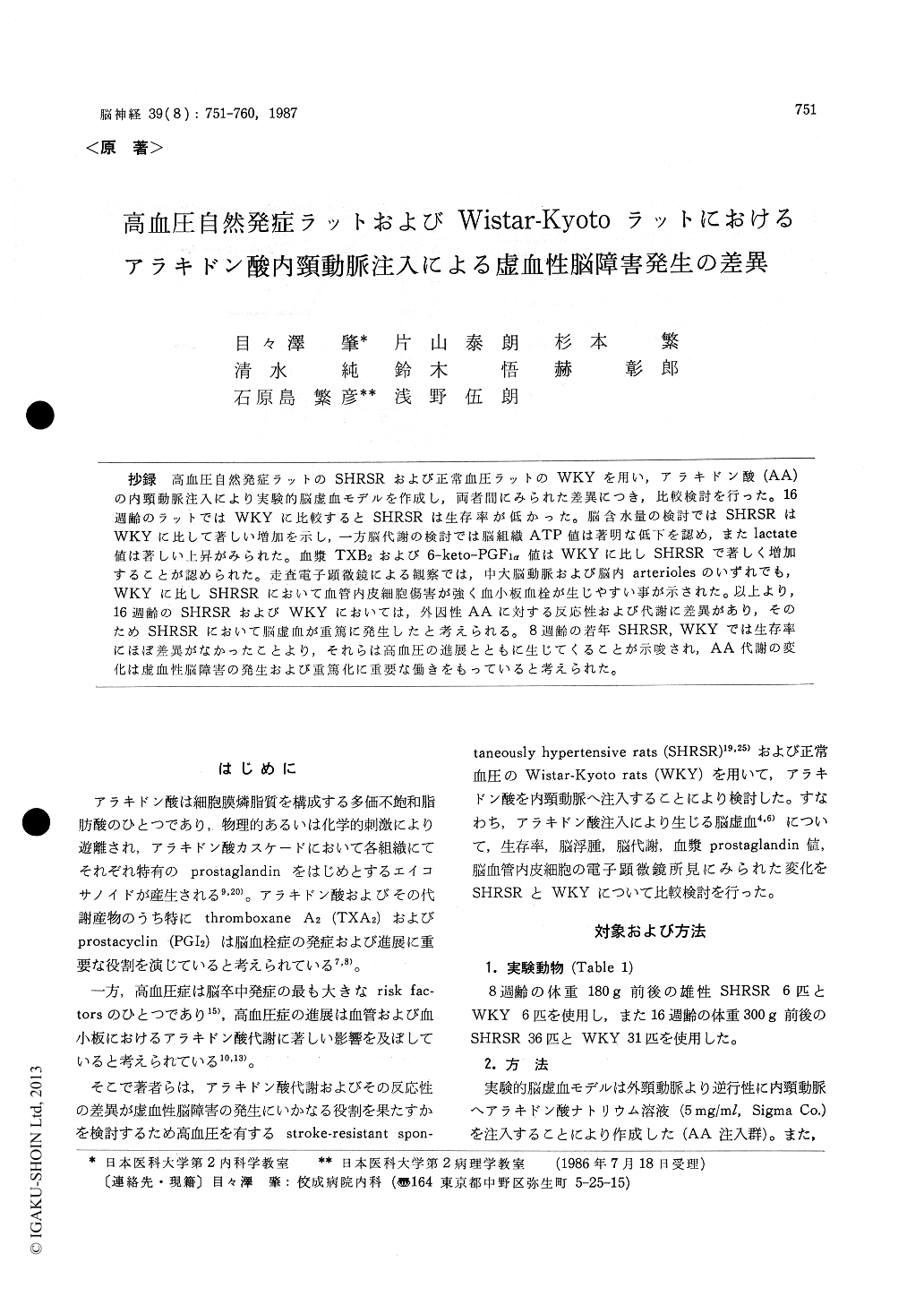
抄録 高血圧自然発症ラットのSHRSRおよび正常血圧ラットのWKYを用い,アラキドン酸(AA)の内頸動脈注入により実験的脳虚血モデルを作成し,両者間にみられた差異につき,比較検討を行った。16週齢のラットではWKYに比較するとSHRSRは生存率が低かった。脳含水量の検討ではSHRSRはWKYに比して著しい増加を示し,一方脳代謝の検討では脳組織ATP値は著明な低下を認め,またlactate値は著しい上昇がみられた。血漿TXB2および6-keto-PGF1α 値はWKYに比しSHRSRで著しく増加することが認められた。走査電子顕微鏡による観察では,中大脳動脈および脳内arteriolesのいずれでも,WKYに比しSHRSRにおいて血管内皮細胞傷害が強く血小板血栓が生じやすい事が示された。以上より,16週齢のSHRSRおよびWKYにおいては,外因性AAに対する反応性および代謝に差異があり,そのためSHRSRにおいて脳虚血が重篤に発生したと考えられる。8週齢の若年SHRSR,WKYでは生存率にほぼ差異がなかったことより,それらは高血圧の進展とともに生じてくることが示唆され,AA代謝の変化は虚血性脳障害の発生および重篤化に重要な働きをもっていると考えられた。
The Second Department of Internal Medicine, Arachidonic acid (AA) is a potent stimulator of platelet aggragation and is known to induce endothelial cell damage and edema in the brain. An ischemic cerebral infarction model can be pro-duced in rats by an internal carotid infusion of AA. In this study, water content, ATP and lactate in the brain and plasma prostaglandins (throm-boxane B2 ; TXB2 and 6-keto-prostaglandin F1α ; 6-keto-PGF1α ) were measured and histological observations were made after an internal carotid infusion of AA in two strains of rats; stroke-resistant spontaneously hypertensive rat (SHRSR) and normotensive Wistar-Kyoto (WKY) rats. It is known that the blood pressure of the SHRSR begins to rise at about 8 weeks old.
Throughout the study, AA was administered into the internal carotid artery under 2% fluothane anesthesia. In the first study, AA (1.7 mg/kg BW) was administered to 8- and 16-week-old rats and the length of survival was observed. In the second study, water content, ATP and lactate in the brain and plasma TXB22 and 6-keto-PGF1α were mea-sured 3 hours after AA (0.85 mg/kg BW) administ-ration. In the third study, rats were perfusion-fixed 3 hours after the AA (1.7 mg/kg BW) infu-sion and cerebral arteries were observed by scan-ning electron microscope (SEM).
During the 6 hour observation period, there was no difference between the two groups in the num-ber of 8-week-old rats which survived. However, the figure became significantly lower in the SHRSR at 16 weeks old. The water content in the bilateral cerebral hemispheres was significant-ly larger in the SHRSR than in the WKY rats. The ATP content of the region was significantly less in the SHRSR than in the WKY rats. The lactate content in the frontal region was signifi-cantly larger in the SHRSR than in the WKY rats, but there were no differences between the two strains in the occipital region. Prior to AA ad-ministration, there were no differences in plasma TXB2 and 6-keto-PGF1α levels of the two strains. After the AA infusion, the magnitude of plasma prostaglandin elevations was significantly larger in the SHRSR than in the WKY rats. Upon SEM examination, the luminal surfaces of the middle cerebral arteries and intracerebral arterioles re-vealed endothelial damage and thrombi formations were more prevalent in the SHRSR than in the WKY rats.
These results show that the AA metabolism and the responses to infused AA in platelets and cerebrovascular endothelial cells differ between SHRSR and WKY rats. These differences may contribute to the severity of cerebral ischemia in SHRSR. The developement of hypertension in SHRSR may be related to the differences in the AA metabolism of the two strains.

Copyright © 1987, Igaku-Shoin Ltd. All rights reserved.


