Japanese
English
- 有料閲覧
- Abstract 文献概要
- 1ページ目 Look Inside
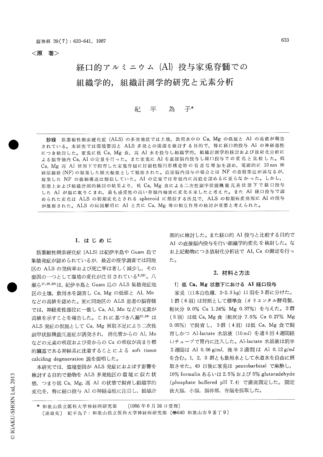
抄録 筋萎縮性側索硬化症(ALS)の多発地区では土壌,飲用水中のCa,Mgの低値とAlの高値が報告されている。本研究では環境要因とALS多発との関連を検討する目的で,特に経口的投与Alの神経毒性につき検討した。家兎に低Ca, Mg食,高AI水を投与し組織学的,組織計測学的検討および放射化分析による脳脊髄内Ca, Alの定量を行った。また家兎にAlを直接脳内投与し経口投与での変化と比較した。低Ca, Mg高Al状態下で飼育した家兎脊髄に好銀性類円形構造物の有意な増加を認め,電顕的に10nm神経原線維(NF)の凝集した腫大軸索として観察された。直接脳内投与の場合とはNFの蓄積部位が異なるが,凝集したNFの微細構造は類似していた。Alの定量では脊髄内に高値を認めるに至らなかった。しかし,形態上および組織計測的検討の結果より,低Ca,Mg食による二次性副甲状腺機能亢進状態下で経口投与したAlが脳に取りこまれ,最も感受性の高い脊髄内軸索に変化を来したと考えた。またAl経口投与で認められた変化はALSの初期変化とされるspheroidに類似する所見で,ALSの初期病変発現にAlの関与が推察された。ALSの病因解明にAlと共にCa, Mg等の相互作用の検討が重要と考えられた。
Environmental factor is noteworthy for patho-genesis of ALS. Yase reported low contents of Ca, Mg and high contents of Al, Mn in envi-ronmental samples (soil and water) obtained from Kii Peninsula and Guam where ALS has been occurring in high incidence.
In this paper, to evaluate the role of these environmental factors for degeneration of ALS, morphological, morphometrical and metal analy-tical studies on experimental animals will be de-scribed with special attention to the neurotoxicity of oral administration of Al.
The rabbits (2-2.3 kg) were divided into 3 groups. Group 1 was fed standard diet (Ca 1.24% Mg 0.37%), Group 2 was fed low-Ca Mg diet (Ca 0.27% Mg 0.05%) for 40 days, and Group 3 was fed low-Ca Mg diet for 40 days and was injected Al-lactate solution (10 ml) into the stomach through the tube 6 times/week for 4 weeks. Al-lactate solution contained Al in dosage of 0.06 g/ml for the first 2 weeks and 0.12 g/ml for the following 2 weeks. And also to compare with the change by oral administration of Al, rabbits were injected Al-phosphate intracerebrally 14 days before the examination.
The rabbits were anesthetized and perfused through the aorta with 10% formalin or 2.5% and 5% glutaraldehyde (pH 7.4), and examined light and electron microscopically. For morphometricalexamination, 100 sections of 6 microne specimen at intervals of 30 micrones in each rabbit spinal cord (c-5) were prepared and examined using the ocular micrometer. Contents of Al and Ca of the spinal cord and cortex were measured by neutron activation analysis with formalin fixed samples.
Morphologically, the argyrophilic bodies were occasionally recognized in the spinal cords of Group 3 and Group 2 rabbits, and rarely in Group 1 rabbits. These structures were significantly in-creased in the Group 3 rabbits (p<0.01). The fine structures of these argyrophilic bodies were recognized as axonal swellings packed with inter-woven, small bundles of 10 nm neurofilaments. The accumulation of the neurofilaments was loca-lized in the axon of the spinal cord.
Al contents in the spinal cords of Group 3 rabbits were not elevated in this study, but Ca contents in the spinal cords and the cortices of Group 2 and Group 3 rabbits were significantly increased (p<0.05).
The fine structure of the neurofilamentous ac-cumulation of Group 3 rabbits was similar with the neurofibrillary changes which was recognized in the rabbits treated with Al-phosphate intra-cerebrally, although there were some differences in the localization of the neurofilament. Based on morphological and morphometrical results, Al by oral administration seemed to have important role for the formation of the axonal swelling under the condition of secondary hyperparathyroidism induced by low-Ca Mg diet.
Spheroid has been regarded as one of the initial changes of ALS. The fine structure of the axonal swelling of Group 3 rabbits was similar with the spheroid of ALS. And so Al might have some role for development of the initial change of ALS. It should be pursued the role of Al with interaction of Ca and Mg for the neurodegene-ration in ALS.

Copyright © 1987, Igaku-Shoin Ltd. All rights reserved.


