Japanese
English
- 有料閲覧
- Abstract 文献概要
- 1ページ目 Look Inside
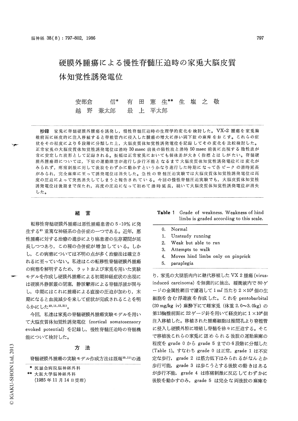
抄録 家兎に脊髄硬膜外腫瘍を誘発し,慢性脊髄圧迫時の生理学的変化を検討した。VX−2腫瘍を家兎胸椎前面に経皮的に注入移植すると脊椎管内に侵入した腫瘍の増大に伴い両下肢の麻痺をおこす。これらの症状をその程度により6段階に分類した上,大脳皮質体知覚性誘発電位を記録してその変化を比較検討した。正常家兎の大脳皮質体知覚性誘発電位は潜時30msec前後の陽性波と潜時50msec前後に出現する陰性波が常に安定した波形として記録される。振幅は正常家兎においても個体差が大きく指標とはしがたい。脊髄硬膜外腫瘍群については,下肢の運動障害が進行し歩行不能となるまで大脳皮質体知覚性誘発電位には変化がみられず,疼痛刺激に対して後肢をわずかに動かすというかなり進行した時期になって各ピークの潜時延長がみられ,完全麻痺に至って誘発電位は消失した。急性の脊髄圧迫実験では大脳皮質体知覚性誘発電位は高度の圧迫によって突然消失してしまうと報告されている。今回の慢性脊髄圧迫実験でも,大脳皮質体知覚性誘発電位は後期まで保たれ,高度の圧迫になって初めて潜時延長,続いて大脳皮質体知覚性誘発電位が消失した。
An experimental model of spinal cord compres-sion was developed in rabbits by epidural neo-plasms which were injected anterior to the T13 vertebral body and grew into the spinal canal through the intervertebral foramina. With this experimental model, the neurological condition of the animals was monitored using a scale and changes of somatosensory evoked potentials (SEPs) were studied to evaluate the neurophysiological effect of experimental chronic cord compression.
The animals were immobilized with pancuro-nium bromide and artificial respiration was main-tained through a tracheostomy. SEPs were recorded by silver ball electrodes which were positioned epidurally over the somatosensory cortex through small burr holes. A subcutaneous needle placed at the nose served as a reference electrode. Right hind paw was stimulated via two percutaneous needles with 0.1 msec rectangular impulses suffi-ciently strong to produce motor responses, ranging from 10 to 20 volt in control rabbits. Electrical stimuli were delivered at a rate of 1 Hz. The intensity of electrical stimulation was raised up to 300 volt, when no consistent SEP was observed in the rabbit with spinal neoplasm. The SEP was summated by averaging 50 successive cortical transients with the analysis time of 200 and 500 msec.
The cortical SEPs in the rabbit normally con-sisted of a positive-negative sequence, which we labelled P1, N1, P2, N2 and so on. Early peaks, P1 and N1, were observed constantly with average latencies of 30.1 and 53.3 msec respectively in normal rabbits. The variability of amplitudes seen even in control animals made them a less useful measure of function than latencies.
Normal SEPs were preserved until the animals demonstrated moderate paraparesis. The rabbits with advanced symptoms showed marked delay in peak latencies of SEPs and SEPs were finally abo-lished in the animals showing complete paraplegia.
It was reported that SEP suddenly disappeared in the animals with acute cord compression. In the present study, we introduced chronic cord compression model and demonstrated that the disturbance of neural conduction gradually devel-oped. And SEP disappeared shortly after latency shift was observed at a final stage.
The experimental model described here appears to be useful in evaluating the pathophysiological change of chronic spinal cord compression.

Copyright © 1986, Igaku-Shoin Ltd. All rights reserved.


