Japanese
English
- 有料閲覧
- Abstract 文献概要
- 1ページ目 Look Inside
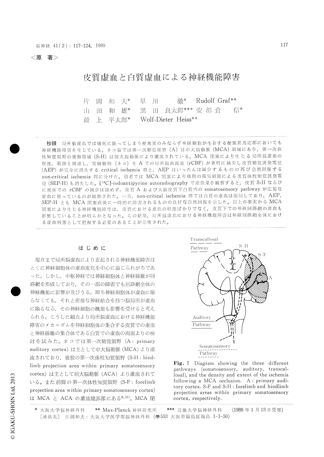
抄録 局所脳虚血では壊死に陥ってしまう梗塞巣のみならず神経組胞が生存する梗塞巣周辺部においても神経機能障害を生じている。ネコ脳では第一次聴覚領野(A)は中大脳動脈(MCA)領域にあり,第一次体性知覚領野の後脚領域(S-H)は前大脳動脈により灌流されている。MCA閉塞により生じる局所脳虚血の程度,範囲と関連し,実験動物(ネコ)をAでの局所脳血流量(rCBF)が著明に減少し皮質聴覚誘発電位(AEP)が完全に消失するcritical ischemia群と,AEPはいったんは減少するものの再び自然回復するnon-critical ischemia群に分けた。前者ではMCA閉塞により後脚の電気刺激による皮質体性知覚誘発電位(SEP-H)も消失した。[14C]-iodoantipyrine autoradiographyで虚血巣を観察すると,皮質S-Hならびに視床でのrCBFの滅少は認めず,皮質Aおよび人脳皮質下白質内のsomatosensory pathwayが広範な虚血に陥っているのが観察された。一方,non-critical ischemia群では白質の虚血は限局しており,AEP,SEP-HともMCA閉塞直後に一時的に障害されるものの良好な自然回復を示した。以上の事実からMCA閉塞により生じる神経機能障害は,皮質における虚血の程度ばかりでなく,皮質下での神経同路網の虚血も影響していることが明らかとなった。この結果,局所脳虚血における神経機能障害は神経回路網全体における虚血病態として把握する必要のあることが示唆された。
We evaluated regional cerebral blood flow (rCBF) by means of hydrogen clearance method as well as [14C]-iodoantipyrine autoradiographic method, cortical auditory evoked potentials (AEP), somatosentory evoked potentials (SEP) induced by forelimb (median nerve) stimulation (SEP-F), and SEP induced by hindlimb (tibial nerve) stim-ulation (SEP-H) in cats after occlusion of the left middle cerebral artery (MCA) under alpha-chloralose anesthesia. According to the degree of ischemia, the experimental animals were divided into two groups. One was the critical ischemia which was defined as permanent total suppression of AEP, and low residual blood flow in the auditory cortex. And the other was the non-critical ischemia which included transient suppression and spontaneous recovery of the cor-tical sensory evoked potentials, and high residual blood flow (>15 ml/100 g/min). In one cat with transient suppression of three kinds of sensoryevoked potentials, the [14C]-iodoantipyrine (IAP) autoradiograph revealed only a limited ischemic area of subcortical white matter. In the critical ischemia group, ischemia of the primary sensory cortex ranged from the mostly affected primary auditory cortex (supplied by the MCA) to the least affected hindlimb projection area within primary somatosensory cortex (supplied by the ACA). The forelimb projection area of the primary somatosensory cortex (supplied by both ACA and MCA) showed a mild or moderate reduction of rCBF after occlusion. The [14C]- IAP autoradiograph showed severe reduction of the white matter including the somatosensory pathway in the wide range. However, rCBF in the thalamus and hindlimb projection area within somatosensory cortex was almost intact in the cat with critical ischemia. These rCBF studies concluded that the suppression of AEPs was at-tributable to cortical ischemia, and the suppres-sion of SEP was attributed to white matter ischemia. We analyzed the acute sequential changes of multimodality evoked potentials (AEP, SEP-F, and SEP-H) using a multiplex stimulation and analysis system. AEPs were abolished within 3 min. after occlusion, whereas the decrease in SEP-Hs started after about 5 minutes, then SEP-Hs disappeared within 10 minutes. According to SEP-F two components of the SEP changes were shown : an early immediate reduction and a later decrease occurring about 5 minutes after occlusion. This time lag possibly reflects the resistance of the white matter to ischemia, compared to cortex.

Copyright © 1989, Igaku-Shoin Ltd. All rights reserved.


