Japanese
English
- 有料閲覧
- Abstract 文献概要
- 1ページ目 Look Inside
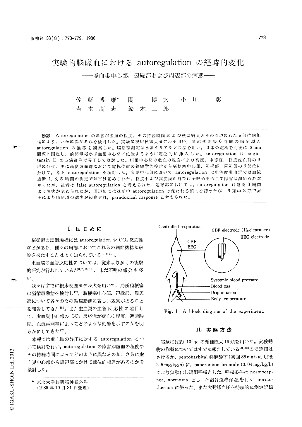
抄録 Autoregulationの障害が虚血の程度,その持続時間および梗塞病巣とその周辺にわたる部位的相違により,いかに異なるかを検討した。実験に視床梗塞犬モデルを用い,血流遮断後6時間の脳循環とautoregulationの推移を観察した。脳循環測定は水素クリアランス法を用い,3本の電極を前後に3mm間隔に固定し,前部電極が虚血巣中心部に位置するように定位的に挿入した。autoregulationはangio-tensin IIの点滴静注で昇圧して検討した。病巣中心部の虚血の程度により高度,中等度,軽度虚血群の3群に分け,更に高度虚血群において電極位置の組織学的検討から脳梗塞中心部,辺縁部,周辺部の3部位に分けて,各々autoregulationを検討した。病巣中心部においてautoregulationは中等度虚血群では血流遮断1,3,5時間の測定で障害は認められた。軽度および高度虚血群では全経過を通じて障害は認められなかったが,後者はfalse autoregulationと考えられた。辺縁部においては,autoregulationは遮断3時間より障害が認められたが,周辺部では遮断中autoregulationは保たれる傾向を認めたが,6頭中2頭で昇圧により脳循環の減少が観察され,paradoxical responseと考えられた。
In order to elucidate the relationship between the degree of autoregulatory loss and the intensity or the duration of ischemia, and the difference of locations in and around the ischemic focus, we used the canine thalamic infarction model and studied the sequential changes of rCBF and auto-regulation during 6 hours following vascular occlusion. The value of rCBF was measured by the hydrogen clearance method and autoregulation was tested by raising the blood pressure with drip infusion of angiotensin II.
In the center of ischemic focus, autoregulation was impaired after 1,3 and 5 hours following occlusion in the animals with moderate ischemia. In the animals with mild and severe ischemia, autoregulation was preserved during occlusion, but in the latter we thought it false autoregulation. In the periphery of infarctic focus with severe ischemia, autoregulation was impaired after 3 and 5 hours following occlusion. Outside of the in-farctic focus with severe ischemia, autoregulation was preserved during occlusion, but in 2 of 6 animals rCBF decreased despite of raising blood pressure and it was thought to be paradoxical response.

Copyright © 1986, Igaku-Shoin Ltd. All rights reserved.


