Japanese
English
- 有料閲覧
- Abstract 文献概要
- 1ページ目 Look Inside
はじめに
脳梗塞については従来よりさまざまな研究が行なわれてきたが,近年,血行再建などの外科治療の発達に伴い,脳梗塞の病態生理について,より詳細な研究がなされるようになつてきた。
このなかで脳梗塞の治療と密接な関係がある脳乏血発現後の神経細胞の経時的変化についての報告は,従来より散見され2,6,7,8,11,17),われわれも局所脳虚血のモデルである視床梗塞モデル犬27)を用いて報告してきた。しかし,これまでの報告は,脳虚血といつても完全虚血であるのか不完全虚血であるのか,また病巣の大小,さらに個休差による病巣の不均一など実験モデルの面からみても種々の問題があり,必ずしも満足のゆくものではなかつた。
Having developed a canine model for complete ischemia of a cerebral hemisphere by means of unilateral occlusion of all the arteries at the trunk of the brain, light and electron microscopic obser-vations on the neurous of the third layer of the cerebral cortex were made following vascular oc-clusion. Similar observations were also made in animals administered mannitol (2g/kg), glycerol (1g/kg) or 25% glucose (10cc/kg) prior to occlusion to determine their effects in preventing cerebral infarction.
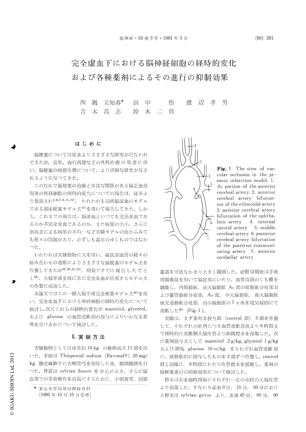
1) For production of infarction, the internal carotid artery, bifurcation of the A1 portion of the anterior cerebral artery and the ophthalmic and ethmoidal arteries, the A2 portion, the middle cerebral artery, bifurcation of the posterior cerebral and posterior communicating arteries, and the anterior cerebellar artery were occluded unilaterally simultaneously in 10kg adult dogs.
2) Samples were obtained from the cerebral cortex at various intervals for 6 hours following occlusion in each dog. Sixty neurons from each sample were studied with the light microscope and neuronal changes classified into 5 categories : Normal, Borderline, Microvacuolation, Shrinkage and Swelling. The percentage of cells showing each change after various periods of occlusion were recorded.
3) The average values from the 5 untreated control animals indicated: 90% of the neurons were Normal or Borderline after 30min occlusion, Shrinkage was first seen after 45min, and Micro-vacuolation was seen in 30% after 60min occlusion. Microvacuolation was found in 40%, Swelling appeared after 120min and Shrinkage accounted for 35% after 180min occlusion. After 240min, no normal cells were seen and Shrinkage accounted for 45%. After 300 and 360min occlusion, Shrink-age or Swelling was seen in roughly 80% of the neurous.
4) Electron microscopically, primarily swelling of mitochondria and rough endothelial reticulum were found in borderline and microvacuolation cells. As the time of cerebral ischemia increased, the electron density of the cytoplasm in cells showing shrinkage increased and destruction of a portion of the cytoplasm was seen in the cells showing swelling.
5) Although slight preventive effect was ob-tained by administration of glycerol or glucose, only mannitol significantly suppressed the develop-ment of cerebral infarction.
Copyright © 1981, Igaku-Shoin Ltd. All rights reserved.