Japanese
English
- 有料閲覧
- Abstract 文献概要
- 1ページ目 Look Inside
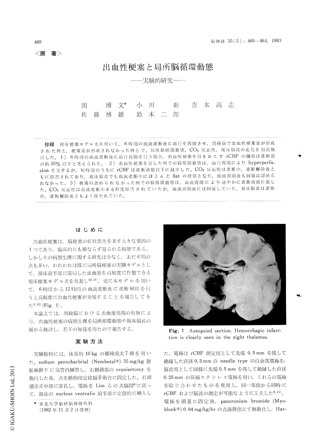
抄録 視床梗塞モデル犬を用いて,6時間の血流遮断後に血行を再開させ,剖検脳で出血性梗塞巣が形成された例と,梗塞巣が形成されなかった例とで,局所脳循環動態,CO2反応性,視床脳波の変化を対比検討した。1)6時間の血流遮断後に血行再開を行う場合,出血性梗塞を引きおこすrCBFの閾値は遮断前の約50%以下と考えられた。2)出血性梗塞を呈した例での脳循環動態は,血行再開によりhyperperfu—sionを呈するが,短時間のうちにrCBFは遮断前値以下に誠少した。CO2反応性は遮断中,遮断解除後ともに障害されており,視床脳波でも血流遮断中にほとんどflatの状態となり,血流再開後も回復は認められなかった。3)梗塞の認められなかった例での脳循環動態は,血流再開により速やかに遮断前値に復した。CO2反応性は血流遮断中ある程度障害されていたが,血流再開後には回復していた。視床脳波は遮断中,遮断解除後ともよく保たれていた。
Using our previously reported "thalamic infarc-tion model in the dogs", it was found that hemorrhagic infarction can be produced at a high frequency following recirculation after 6-12 hours of vascular occlusion. In the present study, in order to elucidate the pathophysiology of hemor-rhagic infarction, we have undertaken a study of the relations among the histological findings, de-gree of ischemia, circulatory dynamics, CO2 re-sponse and EEG findings after vascular occlusion of 6 hours.
In all animals where rCBF was found to fall to less than 50% due to vascular occlusion, hemor-rhagic infarction was found. The hemodynamics of those animals presenting hemorrhagic infarction was such that reflow resulted in a transient in-crease in rCBF followed by decrease within a short period. After a few hours, rCBF values had fallen to pre-occlusion levels. During the 6 hours occlusion, electrical activity became almost flat, and recovery following reflow was not seen. The CO2 response was found to be disturbed imme-diately following vascular occlusion and also did not recover following reflow. In contrast, among the animals in which hemorrhagic foci were not found, reflow resulted in recovery of rCBF to pre-occlusion levels within a short period. Elect-rical activity of the brain and CO2 response were found to be maintained throughout the period of occlusion and thereafter in these animals.

Copyright © 1983, Igaku-Shoin Ltd. All rights reserved.


