Japanese
English
- 有料閲覧
- Abstract 文献概要
- 1ページ目 Look Inside
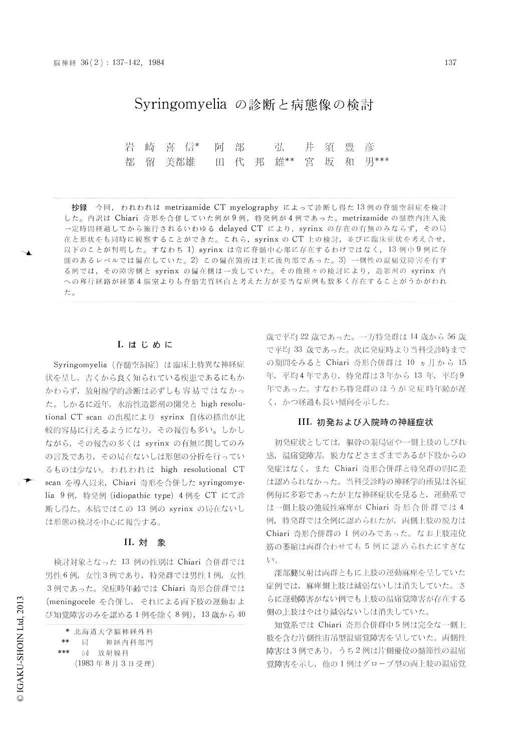
抄録 今回,われわれはmetrizamide CT myelographyによって診断し得た13例の脊髄空洞症を検討した。内訳はChiari奇形を合併していた例が9例,特発例が4例であった。metrizamideの髄腔内注入後一定時間経過してから施行されるいわゆるdelayed CTにより,syrinxの存在の有無のみならず,その局在と形状をも同時に観察することができた。これら,syrinxのCT上の検討,並びに臨床症状を考え合せ,以下のことが判明した。すなわち1) syrinxは常に脊髄中心部に存在するわけではなく,13例中9例に脊髄のあるレベルでは偏在していた。2)この偏在箇所は主に後角部であった。3)一側性の温痛覚障害を有する例では,その障害側とsyrinxの偏在側は一致していた。その他種々の検討により,造影剤のsyrinx内への移行経路が経第4脳室よりも脊髄実質経由と考えた方が妥当な症例も数多く存在することがうかがわれた。
Thirteen cases of syringomyelia which was con-firmed by metrizamide CT myelography (MCTM) were reported.
Nine cases were associated with Chiari malfor-mation, and four were idiopathic type. Delayed CTM which were performed in several hours after the metrizamide myelography disclosed not only the existence of the syrinx but its shape and location.
From the investigation of their clinical findings together with the appearance of their syrinx on CT scan, the following results were obtained.
1) The syrinx was not always situated centrally within the cord, but had laterality at some spinal level in nine out of thirteen cases.
2) The main laterality of the syrinx seemed to be located in the posterior horn.
3) In the cases which had unilateral dissociated sensory disturbance, the laterality of sensory dis-turbance corresponded with the side of syrinx.
These results suggested the possibility that the intrathecal injected contrast materials entered into the syrinx via the dorsolateral sulcus of the cord rather than via IV ventricle in some cases.

Copyright © 1984, Igaku-Shoin Ltd. All rights reserved.


