Japanese
English
- 有料閲覧
- Abstract 文献概要
- 1ページ目 Look Inside
抄録 マウス胎仔・新生仔の中枢神経系に出現するマクロファージをモノクローナル抗マクロファージ抗体を用いた酵素抗体法(labeled avidin-biotin technique)で経時的に検索した。検索期間は胎生10日より生後21日までで,この期間は大脳でのI期後半からII期およびIII期に相当する。脳内マクロファージは胎生14日でまず第4脳室や側脳室の脈絡叢に出現し,それは脈絡叢形成時に,脈絡叢に進入するくも膜下血管系に由来すると考えられた。大脳実質内ではneuroblast形成期であるII期の胎生14日よりmatrixcell layerとmigrating zoneに出現し,胎生19日ではcortical plateにも散在性に分布していた。これら実質内マクロファージは生後徐々に減少し,細胞構築のほぼ完成する生後9日以降にはほとんど認められなかった。大脳より組織発生の遅れる小脳では出生後より主に内顆粒層にマクロファージが、認められた。脳内マクロファージの出現・分布は組織形成の最も盛んなII期にほぼ集中しており,中枢神経系の組織発生と密接に関連していることが示唆された。
It has been controversial whether macrophages from blood circulation which have phagccytic ac-tivity exist in the normal central nervous system (CNS). Based on the data obtained from adult animals, the origin of phagocytes in the brain has variously been attributed to blood monocytes, pericytes and glial elements.
On the other hand, in fetuses and neonates, although it is known that macrophages exist in the CNS, their precise period of appearance and distribution have not been investigated.
In this paper, overall study on localization cf the fetal and neonatal brain macrophages of mice was carried out immunohistochemically using mo-noclonal antibody against macrophage specific marker (Mac-1) and labeled avidin-biotin techni-que. The antibody used in the experiment well recognized blood monocytes and resident macro-phages in various visceral organs both of the fetus-es and adults.
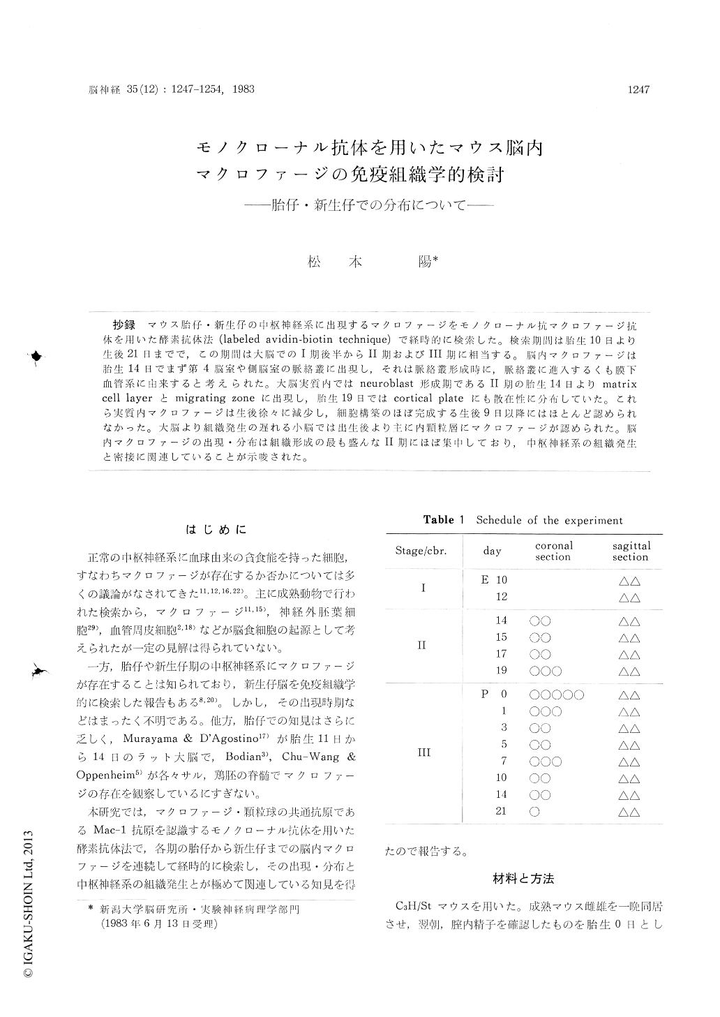
Mice ranging in age from embryonic day 10 (E 10) to postnatal day 21 (P 21) were examined chronologically. The cerebrums of E 10-E 12, E 14-E 19, P 0-P 21 were at the stage of matrix cell, neuroblast, and glioblast formation, respectively.
In the CNS, macrophages first recognized, were located in the choroid plexuses of the fourth and lateral ventricles at E 14. Their number in the choroid plexus of the fourth ventricle increased at E 17-P 3 and gradually decreased thereafter.
Macrophages in the cerebral parenchyme also ap-peared at E 14 in the matrix cell layer and they were detected in the migrating zone at E 15, E 17 and in the cortical plate at E 19. Mapping of the positive cells in the cerebrum at stage II (E 15, E 17, E 19) disclosed the precise distribution of parenchymal macrophages and that macrophages first appeared in the choroid plexus were thought to be derived from the blood vessels which entered the CNS with the choroid plexus.
Almost of all macrophages recognized in the parenchyme of the cerebrum disappeared at P 9 when the cytcarchitecture seemed to be complet-ed. In the cerebellum which formed later than the cerebrum, macrophages appeared after birth and were located mainly in the internal granular layer.
These findings suggested that appearance and distribution of brain macrophages are closely rela-ted to histogenesis of the central nervous system.
Copyright © 1983, Igaku-Shoin Ltd. All rights reserved.