Japanese
English
- 有料閲覧
- Abstract 文献概要
- 1ページ目 Look Inside
抄録 破裂脳動脈瘤によるクモ膜下出血(SAH)後脳浮腫は重大な合併症の一つと考えられ,今回新たな実験的SAH作製モデル,および微量脳組織水分含有量測定法を用いて検討した。調節呼吸下のネコ内頸動脈に糸つき針を刺入,頭蓋閉鎖腔を保った後引き抜きSAHを作製した。頭蓋内圧(ICP),血圧,脳波を3〜24時間観察した。5例で局所脳血流量(rCBF)を放射性マイクロスフィアーにより経時的に4回測定した。全脳60ヵ所の脳組織からrCBFを求め,さらに同組織を真空冷凍乾燥して水分量を求めた。他の5例では著者の改良による脳組織比重測定を行った。SAH後のICP変動は3型に分類できた。Grade I (n=4)では約50mmHgの軽度一過性ICP上昇をみた。Grade II (n=4)では200mmHgに至る高度ICP亢進が10分以上持続した後低下した。rCBFは全脳でSAH前値の20%以下に低下した後血流回復をみた。Grade III(n=2)では高度ICP亢進が持続し脳死に至った。出血の量,広がりとこれらのGradeはよく相関していた。水分量上昇あるいは比重低下はGrade IIにのみ生じ,特に動脈境界領域に著明であった。SAH直後rCBFが20ml/100g/min以下に低下しその後血流同復を示した脳組織が水分量上昇を示した。またSAH後高血圧の持続した例では比量低下が著しく,脳浮腫発生要因の一つであった。
Despite the fact that cerebral edema appears to be a common complication of subarachnoid hemor-rhage (SAH) due to a ruptured cerebral aneurysm, measurements of brain tissue water content have not been carried out in this entity. For this reason, the development of cerebral edema has been analized in a newly developed experimental SAH model.
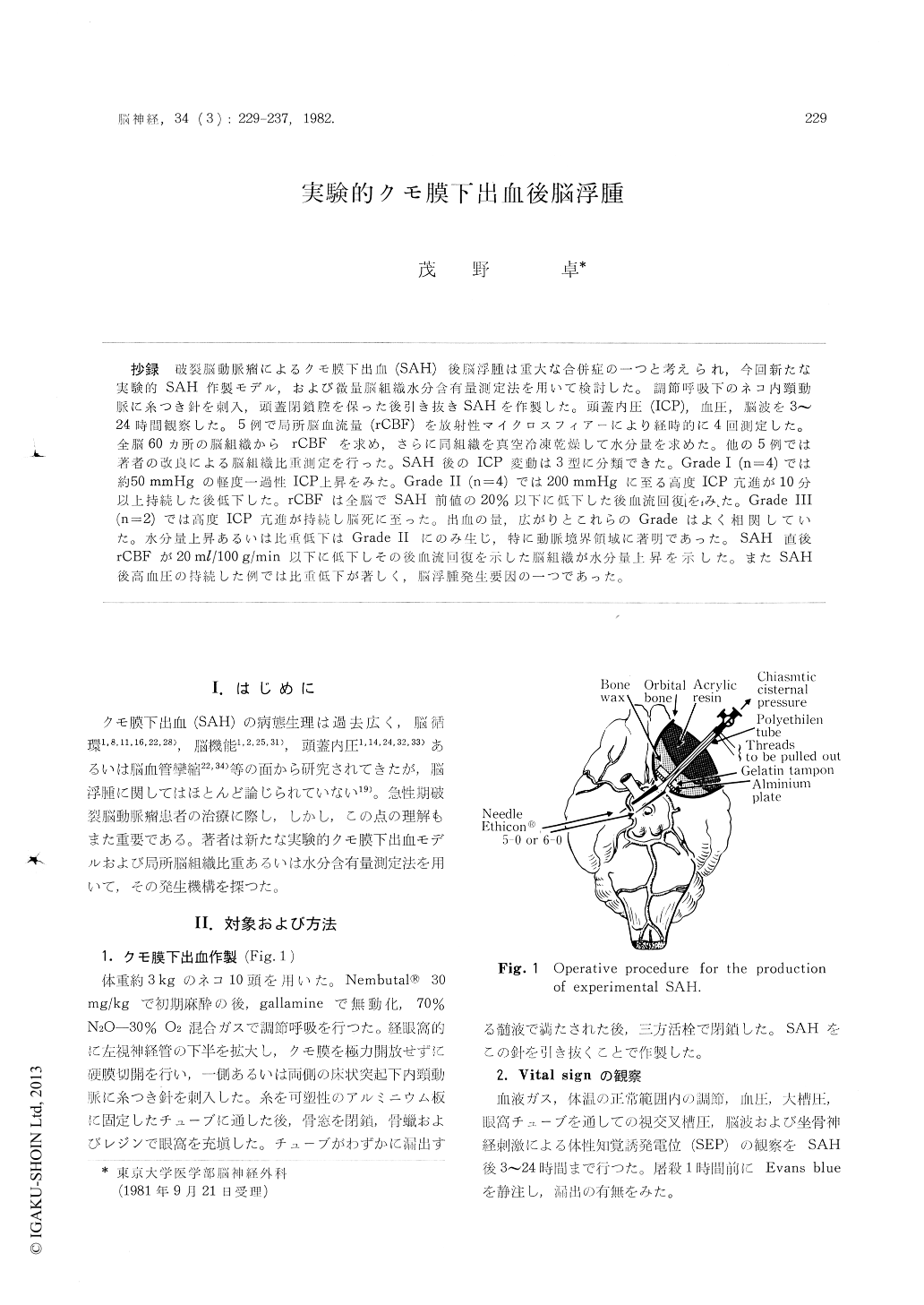
SAH was induced in cats under controled ven-tilation by withdrawing needles previously pierced into one or both infraclinoid internal carotid arteries through a unilateral transorbital approach. Intracranial pressure (ICP), blood pressure, cortical EEG and somatosensory evoked potentials (SEP) to sciatic nerve stimulation were monitored for 3 to 24 hours. Using labelled microspheres of 15 it diameter, four serial determinations of regional cerebral blood flow (rCBF) were performed in one group of five animals before and immediately after SAH as well as 10 to 20 min, and 3 to 6 h post SAH. RCBF were obtained in 60 areas of the brain from tissue samples weighing about 150 mg. Brain tissue water content was determined by the vacuum freeze-drying weighing procedure in the same brain samples. In another group of five ani-mals, brain tissue specific gravity (SG) was measured by microgravimetry in samples of brain tissue weighing 30 to 50 mg. Regional differences in SG as well as in water content were calculated from 21 untreated control animals.
The course of ICP after SAH could be classified into three grades according to the severity of SAH. In grade I (N=4), the increase of ICP was transi-ent and minor up to 50 mmHg without significant effect on vital signs and rCBF. In grade II (N=4), ICP increased up to 200 mmHg with a vasopressor response and total supression of EEG and SEP. RCBF decreased markedly below 20% of control in both cerebral hemispheres, while the brain stem circulation was relatively preserved. However, the distribution of decrease in rCBF showed inhomo-geneous patchy pattern. Ten to 20 min later along with the reduction of ICP, rCBF increased over control, indicating reactive hyperemia. Three to 6 hours later, a great reduction of rCBF was again observed. Extravasation of Evans blue was some-times observed in the parasagittal water-shed areas of the hypertensive animals after SAH. In grade III (N=2), rCBF was sustained at essentially zero flow with the presence of continuously increased ICP above 100 mmHg. Cerebral edema defined as a decrease in SG or an increase in water content was observed in all animals of grade II. When rCBF and brain water content were measured sim-ultaneously, there was a close relationship between the increase in water content and the decrease in rCBF below 20 ml/100 g/min immediately after SAH. In contrast, cerebral edema did not develop in both grade I and grade III animals.
These data suggests that cerebral edema compli-cating SAH is caused by the combination of an initially induced global cerebral ischemia and the subsequent recovery of cerebral circulation. Post SAH hypertension is another factor to exacerbate the development of cerebral edema.
Copyright © 1982, Igaku-Shoin Ltd. All rights reserved.