Japanese
English
- 有料閲覧
- Abstract 文献概要
- 1ページ目 Look Inside
I.はじめに
錐体路は中枢神経系内における主要神経路の一つであり,その起始および迷走束の径路については変性実験,あるいはHRP法による報告,また,その径路については神経線維の数の計測および比較解剖学的記載などがある2,3,7〜9,11〜17,20〜22,25〜27,30,33,34)にもかかわらず,錐体交叉の発生についての記載ははなはだ少ない10,12,13,15,18)。著者は視〔神経〕交叉の形成機序を考察し,その結果に基づいて錐体交叉および文献上見られる錐休交叉の異常例の形成機序についても考察した28,29)。
錐体交叉の交叉後の走行をネコ胎児の脳の標本を用いて観察し,この走行がいかにして決まるかを個体発生学的に考察する。
The course of the pyramidal fibers after decus-sation was investigated in cat fetuses, which were 9-100mm in C. R. length. Every fourth section of sagittal or horizontal serial paraffin sections cut at 10μm was stained after Masson-Goldner's method and impregnated after Bodian's or Loots-Loots-Joubert's method.
The results were summarized as follows:
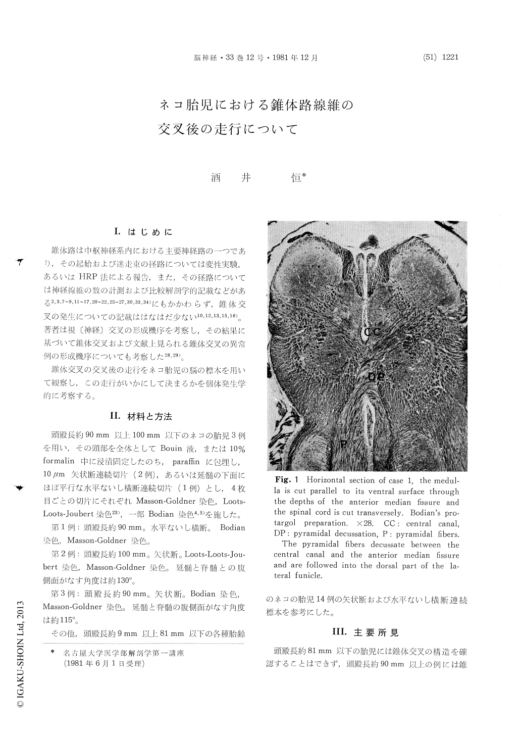
1. The pyramidal decussation was completed infetuses over 90mm in C. R. length between the central canal and the anterior median fissure al-though not demonstrable in those less than 81 mm in C. R. length.
2. In the transverse sections of the spinal cord the crossed pyramidal fibers obliquely extended drawing a slight arc into the dorsal part of the laternal funicle on the opposite side.
3. In the parasagittal sections of the spinal cord the crossed pyramidal fibers fanned out from the pyramidal decussation. The uppermost fibers of them passed dorsolateralward in the same neural segment, in which the decussation occurs, while the lowest fibers dorsocaudalward into the lower segments.
4. The long processes of the ependymal cells of the central canal reached the margin of the anterior median fissure. At the cervical flexure of the brain-stem at the junction of the brain and spinal cord they converged fanwise. The pyramidal fibers, which descended on the ventral surface of the brain-stem to the cervical flexure, crossed here and fanned out in small bundles along and among these ependymal processes dorsocaudalward. There-fore the author holds that these ependymal pro-cesses play a guide-fiber for the crossing pyramidal fibers.

Copyright © 1981, Igaku-Shoin Ltd. All rights reserved.


