Japanese
English
- 有料閲覧
- Abstract 文献概要
- 1ページ目 Look Inside
I.はじめに
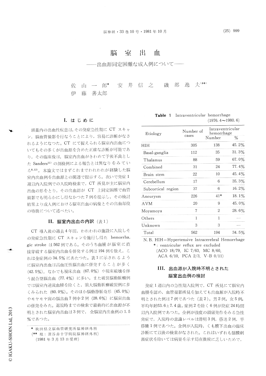
頭蓋内の出血性疾患は,その発症急性期にCTスキャン,脳血管撮影を行なうことにより,容易に診断がなされるようになつた。CTにて捉えられる脳室内出血についてもその多くが出血源を含めた正確な診断が可能であり,その臨床像は,脳室内出血がきわめて予後不良としたSanders11)の剖検例による報告とは異なりをみている8,12)。本論文ではまずこれまでわれわれが経験した脳室内出血例を出血源との関連で提示する。次いで発症1週以内入院例での入院時検索で,CT所見が主に脳室内出血の形をとり,その出血源がCT上同定困難で血管撮影でも明らかにし得なかつた7例を提示し,その検討結果より成人例における脳室出血の病像とその出血部位の特徴について述べたい。
Since Sanders reported the Intraventricular Hemorrhage (IVH) with his autopsied cases in 1881, IVH had assumed to be most serious and fetal condition irrespective of its hemorrhagic source. But after CT scan was applied to such cases, the understanding of IVH has been gradually changed.
We had experienced 194 cases with intraventri-cular hemorrhage among 562 cases with acute hemorrhagic stroke during recent four years in our institute.
Hypertensive intracerebral hemorrhage, especiallythalamic hemorrhage and so-called, combined type hemorrhage which involved the thalamic region, were the commonest origin of these IVH. As for the IVH following ruptured aneurysms, anterior cerebral artery aneurysms were accompanied by it most.
Out of the above 194 cases, those of which were failed to detect their bleeding sites at the first in-vestigation on admission, were seven. For the purpose of elucidating the clinical entity of IVH in adult cases, and the significance of those bleed-ing points, we analyzed these seven cases and tried to find out their hemorrhagic source mainly by further follow-up studies in the time of the discharge.
With follow-up studies, which included one autopsy, four cases were revealed their hemorrhagic source. Thalamus, caudate head, and cerebellar vermis were those of them. Another one, which was shown by autopsy, was small arterio-venous malformation in the splenium of corpus callosum. To detect those regions except the latest one, enhanced CT studies one or two weeks later were most eligible, which disclosed intracerebral resolving hematomas readily. The remaining three cases could not be shown their bleeding sites in spite of applying this methods.
Those seven cases had minimal or no lateralizing signs nor symptoms, but some of them showed limitations of extraocular movement and unequal pupils due to brain stem compression by their hematomas. With surgical intervention, such as continuous ventricular drainage, all except one with AVM were good results at discharge.
Primary Ventricular Hemorrhage (PVH), which is usually seen in preterm infants, is rare in adults. Choroid plexus papilloma or angioma is one of causes in PVH. But this is almost exceptional. The other one is hemorrhage from the ventricular wall or periventricular zone, which are supplied by the centrifugal arterioles derived from choroidal arteries and lenticulostriate arteries (Van den Bergh, 1969). Those angioarchitecture is assumed to be vulnerable to the ischemic condition. Peri-ventricular nuclei, such as thalamus and caudate have many miliary aneurysms, which are considered to be causal pathogenesis of hypertensive intrace-rebral hemorrhage. In IVH with minimal or no damages of cerebral parenchyma as these seven cases, hemorrhagic sources were mainly attributed to these periventricular nuclei which were also supplied by the centrifugal arterioles on their ventricular surface.
Hemorrhagic mechanisms from them might be closely related to their blood supply and this vulnerable angioarchitecture.
Almost all IVH without evident hemorrhagic source by CT scan were secondary hemorrhage from the small lesion of periventricular nuclei in our cases.
Minimal lateralizing signs and symptoms, good results with early surgical procedure was their ordinary profile.

Copyright © 1981, Igaku-Shoin Ltd. All rights reserved.


