Japanese
English
- 有料閲覧
- Abstract 文献概要
- 1ページ目 Look Inside
I.はじめに
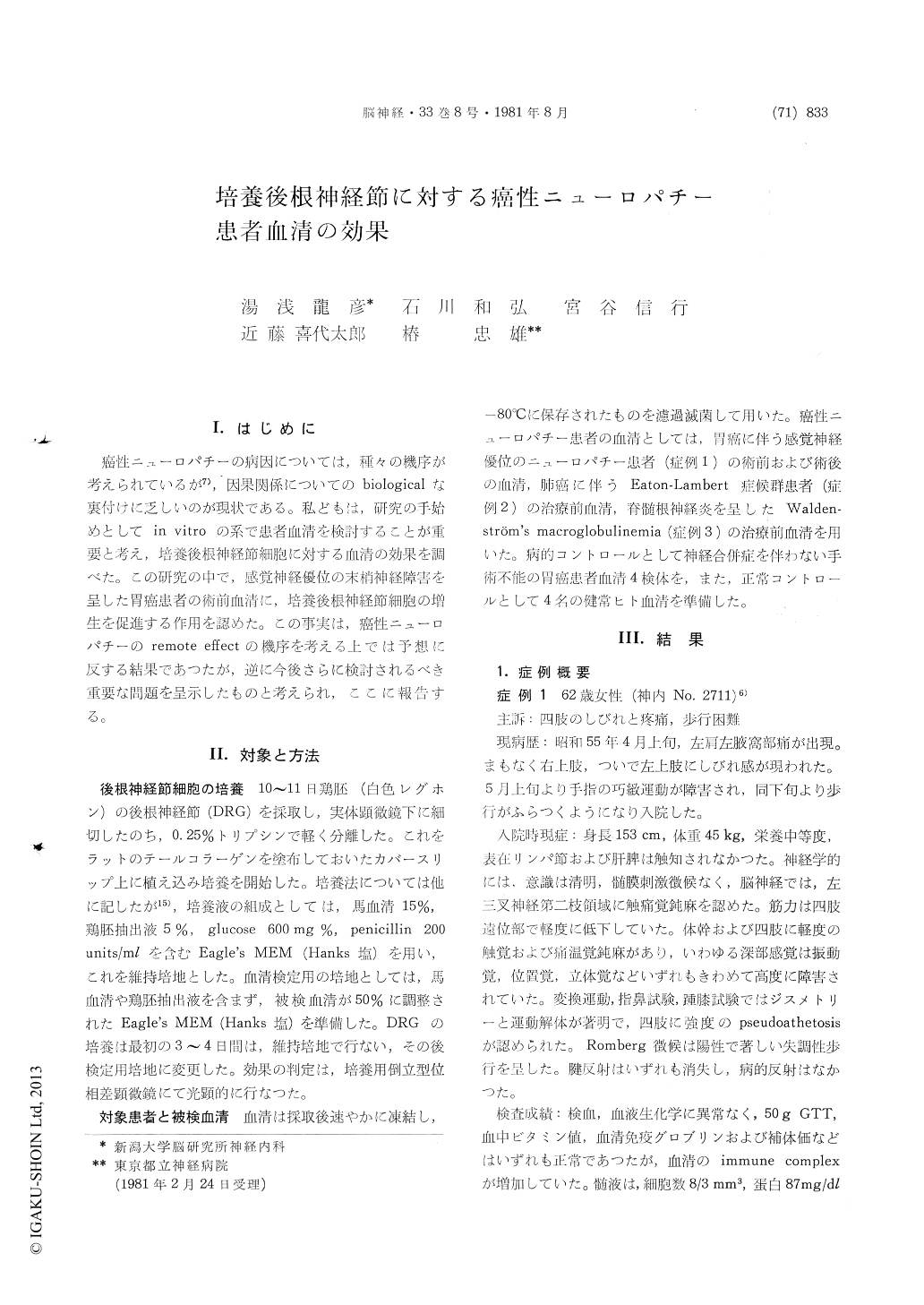
糖性ニューロパチーの病因については,種々の機序が考えられているが7),因果関係についてのbiologicalな裏付けに乏しいのが現状である。私どもは,研究の手始めとしてin vitroの系で患者血清を検討することが重要と考え、培養後根神経節細胞に対する血清の効果を調べた。この研究の中で,感覚神経優位の末梢神経障害を呈した胃癌患者の術前血清に,培養後根神経節細胞の増生を促進する作用を認めた。この事実は,癌性ニューロパチーのremote effectの機序を考える上では予想に反する結果であつたが,逆に今後さらに検討されるべき重要な問題を呈示したものと考えられ,ここに報告する。
Cell-biological effects of sera from patients with carcinomatous neuropathy on cultivated dorsal root ganglia cells were examined.
Dorsal root ganglia, dissected from 10 to 11-day old chick embryos, were partially dissociated by gentle trypsinization, and the explants were placed on collagen-coated cover slips. All cultures were incubated in a humidified atmosphere containg 3% CO2 at 37℃. The culture medium (complete medi-um) was constituted 80% of Eagle's MEM (Hanks base), 15% of horse serum, 5% chick embryo extract, 600mg % of glucose and 200units/ml of penicillin. During the first 3-4 days the cultures were maintained in the complete medium and then the cover slips were placed in an experimental medium with the composition of 50% of experi-mental serum in Eagle's MEM. The experimental sera were obtained from three patients with non-metastatic carcinomatous neuropathy, four diseased control subjects suffering from stomach cancer without any symptom of neurological complications, and four healthy subjects.
The clinical features of the three patients with carcinomatous neuropathy are given briefly ; Case 1: A 62 year-old woman was examined because of pain and dysesthesia of the extremities and difficulty in walking for two months duration. Neurological examination disclosed that she had a sensory dominant neuropathy with severely incoordinated movement and marked pseudoathetosis in the ex-tremities. A gastrectomy was performed because of gastric cancer. There was no evidence of me-tastasis of cancer cells in regional lymph nodes. Her neurological sympoms gradually began to im-prove after the operation. An investigation showed that immune complex was highly positive in the preoperative serum. The sera, for the cultivation of dorsal root ganglia, were obtained before andafter the operation. Case 2: A 52 year-old man was admitted because of fatigability and weakness of the extremities. The chest film revealed an abnormal shadow in the left lung and a cytologic test disclosed atypical cells suggestive of small cell carcinoma of the lung. A typical waxing phe-nomenon was observed on repetitive electric stimulation of the ulner nerve. He was diagnosed as having Eaton-Lambert syndrome associated with lung cancer. A serum was obtained before irradia-tion therapy of the tumor. Case 3: A 65 year-old man had symptoms of motor and sensory distur-bances in the extremities for three months duration. Physical examination disclosed that he had lym-phadenopathy and splenomegaly with slight anemia. Laboratory findings including elevated IgM level in the serum and histopathologic findings of lymph nodes were compatible with the diagnosis of Waldenström's macroglobulinemia. A sample of serum was obtained before the start of therapy.
The dorsal root ganglia cells cultivated in the sera of case 1 showed peculiar effects on the out-growth of neurites and the maturation of cytoplasm: excellent outgrowth was shown when the neurous were cultivated in the serum obtained before the operation. On the other hand, the cells were shrunken in the serum obtained postoperatively. The same excellent results were only shown in the cells cultivated in the complete medium. The sera from two other patients with carcinomatous neu-ropathy and the control subjects showed cytopathic effects on the dorsal root ganglia cells.
These results indicate that the cytopathic factors in the serum of normal subjects should be investi-gated, and we found that these factors were inac-tivated by an incubation at 56℃ for 30min. Also the growth promoting effects seen in the preopera-tive serum of case 1 indicated that there were nerve growth factor (NGF)-like activities in the serum because the culture system adopted here has been used as a biological assay system for NGF. These findings indicate possibilities such as follows: (1) NGF or NGF-like substance might had been secreted from the stomach cancer of case 1, (2) the stomach cancer stimulated the secretion of intrinsic NGF from the NGF secreting organs or tissues, or (3) some specific injury of the nervou system (like dorsal root ganglia lesions in case 1) may promoted the secretion of intrinsic NGF.

Copyright © 1981, Igaku-Shoin Ltd. All rights reserved.


