Japanese
English
- 有料閲覧
- Abstract 文献概要
- 1ページ目 Look Inside
I.はじめに
急性期頭部外傷で,造影剤を用いたCTscanは,時間の浪費の割に得られる情報が少ない,また,当直の脳外科医がたつた独りで重症の頭部外傷患者の診療に当たらねばならないといつた実際の臨床においては,精神的,能力的余裕のないことが多い,などの理由で,従来,造影剤注入(Contrast Enhancement以下CE)によるscanが比較的行なわれてこなかつた傾向にある。したがつて頭部外傷急性期に焦点をあてた,CE症例の報告も少ない13,14)。そこで,急性期頭部外傷患者のCEを用いたCTscanから,実際いかなる情報が得られるかの検討を試みた。
The authors have attempted to do analysis of computerized tomographic scans (CT scans) with contrast enhancement by intravenous contrast medium administration in acute head injuries.
Between April, 1977 and October, 1979, 244 cases of head injuries admitted to the Department of Critical Care Medicine, Nippon Medical School, Sendagi, Tokyo. Out of them 95 patients were chosen because they were all examined with both noncontrast and contrastenhanced CT scans and suffered from (1) acute epidural hematoma, (2) acute subdural hematoma, (3) cerebral contusion, (4) traumatic intracerebral hematoma and/or (5) traumatic intraventricular hemorrhage. 149 cases were excluded from analysis because no such abnormal findings as mentioned above were found or contrast-enhanced CT scans were not performed, or because of contamination of artifacts.
The CT scanner was EMI 1010 and contrast enhancement was performed by intravenous injec-tion of 50 ml of meglumine amidotrizoate (65% AngiografinR) in adults or of 1 ml/kg weight of the same medium in children.
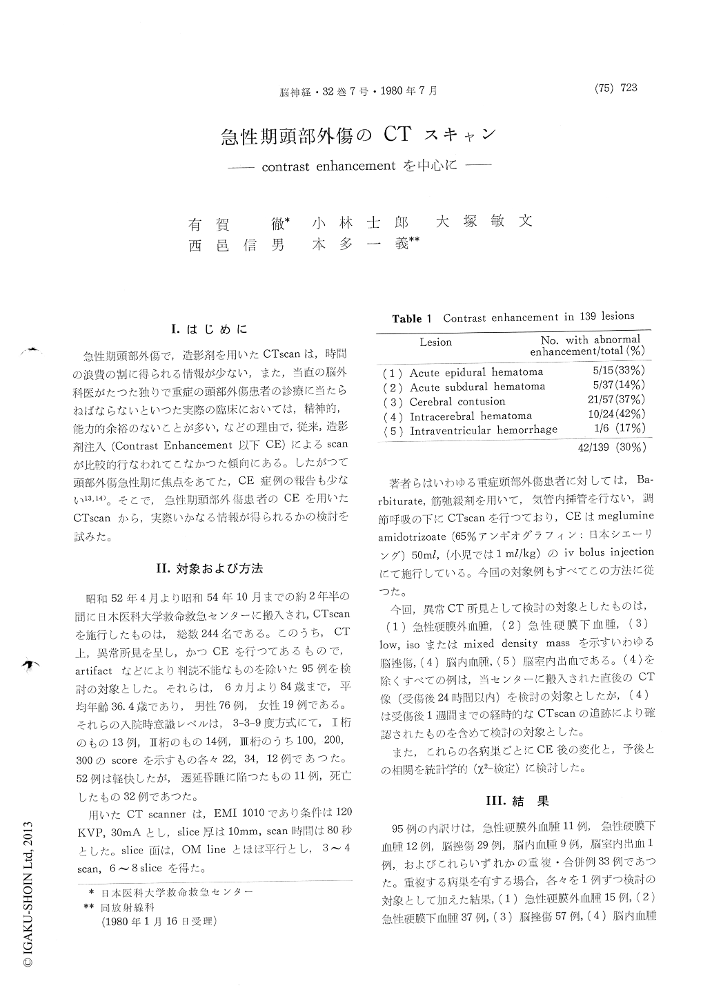
As each of the 95 patients had one or more lesions of (1) to (5), the total number of analyzed lesions was 139 which consisted of (1) 15 cases ofacute epidural hematoma, (2) 37 cases of acute subdural hematoma, (3) 57 cases of cerebral contu-sion, (4) 24 cases of intracebral hematoma and (5) 6 cases of intraventricular hemorrhage.
These lesions were observed within 24 hours post trauma except for traumatic intracerebral hematoma which proves on clinical and experimental analyses to be formed in delayed fashion, closely related to cerebral contusion, the study of which was pursued on serial CT scans performed by the seventh day after trauma.
In 5 out of 15 cases (33%) of acute epidural hema-toma contrast enhancement was seen in the inside and possibly along the internal margin of the hematoma. The latter type of enhancement was revealed in case of relatively large epidural hema-toma and apparently related to plenty of minute bleeding spots on the dural surface which were observed at surgery. Cortical contusion was not verified in such cases. In case of acute subdural hematoma which is mostly accompanied by under-lying cortical contusion, no evidence of enhance-ment along the inner surface of the hematoma was noted. Only 5 out of 37 cases (14%) of acute subdural hematoma demonstated abnormal enhance-ment in the inside of the hematoma.
Cerebral contusion defined by low, iso or mixed density mass lesion on computerized tomography showed two kinds of abnormal enhancement. One of them was that enhancement of contused paren-chyma which was seen usually in low density contused brain. This type of enhancement would reflect leakage of fluid and cell component from damaged vascular endothelium, which is supposed to be etiological for evolution of traumatic intra-cerebral hematoma (contusional hematoma). In fact this enhanced lesion resulted in discovery of evolu-tion of contusional hematoma on serial CT scans.
The other type of enhancement was observed on the cortical sulci where traumatic subarachnoid hemorrhage was evident on noncontrast CT. Just as in acute phase of Reye's syndrome or in other disorders with pathological cerebral vasodilatation, striking enhancement of cortical sulci with subara-chnoid bleeding was demonstrated. The picture presented by these CT scans is compatible with vasoparalysis and petechial bleeding in such areas. Cerebral contusion demonstrated such abnormal enhancement in 21 out of 57 cases (37%).
Among 24 cases of traumatic intracerebral hema-toma, 10 cases (42%) showed linear or dotted enha-ncement in surrounding low density area. Whether this enhancement is due to hyperemia or due to resolving of hematoma remains still unknown.
Only one of 6 cases (17%) of intraventricular hemorrhage showed abnormal enhancement alongthe lateral ventricular wall, probablly reflecting injuries to the ependymal or subependymal layer.
No statistically significant relationship between abnormal enhancement and prognosis in any kind of these lesions was obtained.
Judging from the authors' series of acute head injury patients, the differential diagnosis in indi-visual case would not be difficult without contrast-enhanced CT scans, but pathophysiological changes occurring in acute phase of head injury, especially in cerebral contusion could be manifested far better on computerized tomography with intravenous injection of contrast medium.
Copyright © 1980, Igaku-Shoin Ltd. All rights reserved.