Japanese
English
- 有料閲覧
- Abstract 文献概要
- 1ページ目 Look Inside
I.はじめに
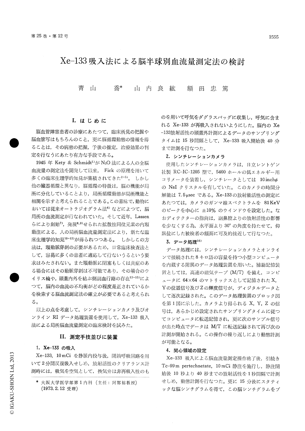
脳血管障害患者の診療にあたつて,臨床所見の把握や脳血管写はもちろんのこと,更に脳循環動態の情報を得ることは,その病態の把握,予後の推定,治療効果の判定を行なうにあたり有力な手段である。
1945年Kety & Schmidt1)がN20法による人の全脳血流量の測定法を開発して以来,Fickの原理を用いて多くの臨床生理学的知見が集積されてきた2〜5)。しかし他の臓器循環と異なり,脳循環の特徴は,脳の機能が局所に分化していることより,局所循環動態が局所機能と相関を示すと考えられることである。この意味で,動物においては従来オートラジオグラム法6)などによつて,脳局所の血流測定が行なわれていた。そして近年,Lassenらにより創始7),発展8,9)せられた拡散性同位元素の内頸動注による,人の局所脳血流量測定法により,新たな臨床生理学的知見10〜12)が得られつつある。 しかしこの方法は,頸動脈穿刺の必要があるため,日常臨床検査法として,容易に多くの患者に適応して行ないうるという要求はみたされない。また頸動脈に閉塞もしくは炎症のある場合にはその動脈穿刺は不可能であり,その場合のウイリス輪や,頭蓋内外を結ぶ側副血行路の存在13〜15)によつて,脳内の血流の不均衡がどの程度是正されているかを検索する脳血流測定法の確立が必要であると考えられる。
It is very important to measure the cerebral blood flow (CBF), which have made for a better understanding of the pathophysiology, prognosis and treatment. In 1963 Lassen and his cooporates reported the quantitative evaluation of r-CBF in man by the use of Kr-85 and multiple external detectors. Their procedures, however, have not become routine examination because carotid arterial puncture is inevitable.
In this work, CBF measurement by the inhala-tion method of Xe-133 using a scintillation cameracombined with an on-line minicomputor was evalu-ated. The patient lied in the supine position and the detector of scintillation camera was placed closely on the frontal plane from anterior 30° axial position. After 10 mCi of Xe-133 was inhaled by means of the closed circuit system for 2 minutes, room air was inspired with non-rebreathing valve. Simultaneously radioactive clearance curve from the head was measured for 40 minutes. The serial R. I. data were transfered to the magnetic tape through the computor having 8K words memory according to the afore-settled sampling time. After the examination of Xe-133 inhalation method the brain scintigram of Tc-99 m administered intra-venously was taken in order to determine the area of interest (AOI). The Xe-133 clearance curve from the AOI was analysed by the method of three compartment analysis which provided the separated blood flows for gray matter, white matter and a slow third component. The third component was presumably due to extracranial tissue.
Hemispheric cerebral blood flow (H-CBF) were measured on 6 patients with cerebrovascular disease, 2 patients with aortic arch syndrome and a mild hypertensive. In these patients no significant dif-ferences were obtained between each H-CBF values except one case.
A patient with the significant low value of the left H-CBF was induced dizziness and syncope by compression of the left carotid artery. The left posterior cerebral artery and communicating artery were demonstrated by the right carotid angiogram. In an aortic arch syndrome patient with remark-able narrowing of left carotid and vertebral arteries and markedly developed collateral vessels, left H-CBF value did not decreased. Both "arm to retina circulation time" using fluolescein and "brain mean transit time" by Tc-99 m showed no signi-ficant differences between each hemispheres cor-responding to that of H-CBF values.
It was concluded that the Xe-133 clearance curve obtained by this procedures provide the information of hemispheric cerebral blood flow, and also this inhalation method had advantages of little load to patient and of application to the patients with obstructive carotid artery. Further studies should be done as routine examination of cerebral bood flow measurement.

Copyright © 1973, Igaku-Shoin Ltd. All rights reserved.


