Japanese
English
- 有料閲覧
- Abstract 文献概要
- 1ページ目 Look Inside
I.はじめに
脊髄前根は運動性であり,後根は知覚性であることは,Bell-Magendieの法則として知られている。しかし,脊髄前根に求心性知覚性線維がある程度含まれていることは,形態学的12,36〜38),電気生理学的4,12,13,25)研究からかなり解明されている。一方,後根に遠心性運動性線維が存在するという報告も古くから見られる14,30,32)。特に,呉,沖中とそのグループは,我国における自律神経学の黎明期ともいえる1930年前後にこの問題に関する広汎な研究を行ない,脊髄後根には多数の遠心性運動性線維が存在し,脊髄副交感神経系を形成するとした16〜19)。しかし,脊髄後根に遠心性線維が存在することに否定的な報告も多数見られ1,9,21,29),この是非についての疑問は決定的解決を見ないまま,久しく新たな進展は見られなかつた。ところが,最近になつて,竈顕レベルで後根切断実験を行ない,腰髄後根には有髄のみならず無髄遠心性線維も存在するとする報告もあり8,27),この問題に対して,新しい視点が提供された。
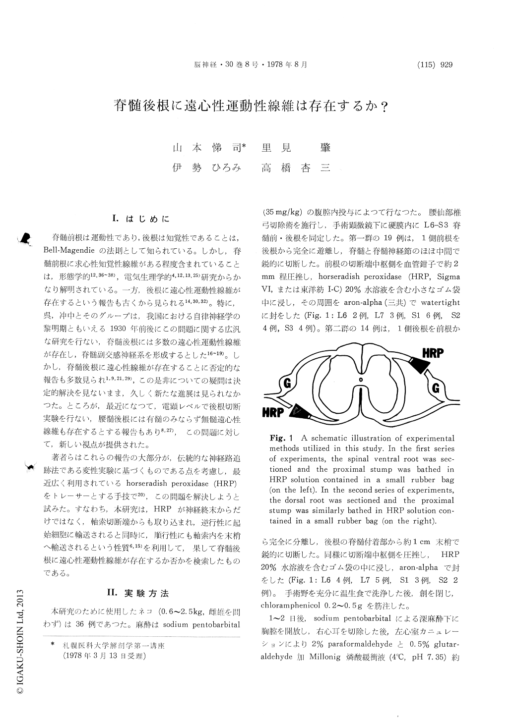
著者らはこれらの報告の大部分が,伝統的な神経路追跡法である変性実験に基づくものである点を考慮し,最近広く利用されているhorseradish peroxidase (HRP)をトレーサーとする手技で20),この問題を解決しようと試みた。
There has been an unsolved question as to whether there are any efferent nerve components in the spinal dorsal roots. Many experimental studies have been done on this subject in attempts to prove or deny the existence of dorsal root efferents. In this study, we investigated this problem in the cat utilizing orthograde as well as retrograde transport of horseradish peroxidase (HRP) through the tran-sected axonal stump. A lumbosacral ventral or dorsal root was identified intradurally in thirty-three cats and the root was sharply severed. The proximal stump of the dorsal or ventral root was bathed in a 20% HRP solution contained in a small rubber bag and the bag was made watertiget with aron-alpha. One or two days later, the animalswere sacrificed by transcardial perfusion of a para-formaldehyde-glutaraldehyde mixture. The next day, the lumbosacral spinal cord was sectioned on a freezing microtome and diaminobenzidine histo-chemistry was performed to visualize HRP reaction products.
1) When the ventral root was bathed, many retrogradely labeled neurons were observed in the somatic motoneuron pools, the cell group X of Onuf, and in the nucleus intermediolateralis inferior, all ipsilaterally to the bathed root.
2) When the dorsal root was bathed, numerous orthogradely labeled primary afferent fibers were identified within the spinal cord. They appearently showed terminal fields in the nucleus proprius and the base of the dorsal horn. Some of them were identified beyond the dorsal horn, particularly in the ventral horn. However, no retrogradely labeled neurons were identified in the spinal cord throughout this series of experiment.
3) As an additional experiment, L7-S2 dorsal and ventral roots were cut in three cats and after four or five days of survival, the spinal cord was fixed and then impregnated by the Fink-Heimer method for degenerating axons. This study revealed es-sentially similar patterns of distribution of primary afferent fibers and their segmental terminal field to cases of dorsal root bathings with HRP.
From the above results, we have concluded that there are no efferent neurons in the lumbosacral spinal cord which give rise to the dorsal root ef-ferents. This implies, in turn, that there are no efferent motor components in the dorsal roots in the lumbosacral cord level in the cat.

Copyright © 1978, Igaku-Shoin Ltd. All rights reserved.


