Japanese
English
- 有料閲覧
- Abstract 文献概要
- 1ページ目 Look Inside
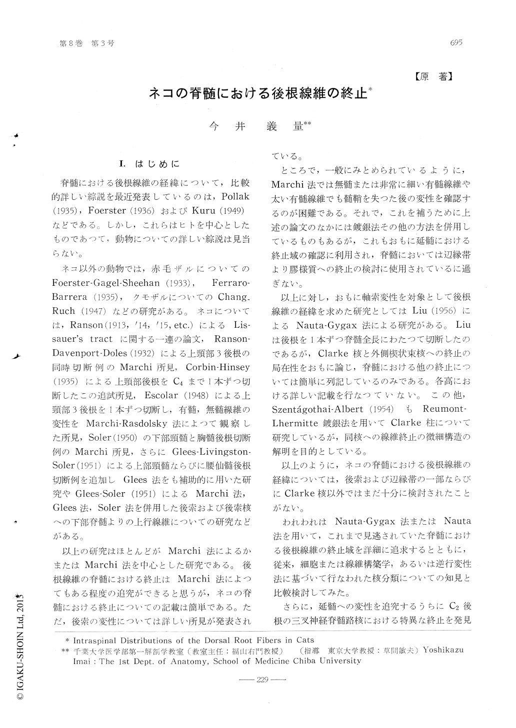
I.はじめに
脊髄における後根線維の経緯について,比較的詳しい綜説を最近発表しているのは,Pollak(1935),Foerster(1936)およびKuru(1949)などである。しかし,これらはヒトを中心としたものであつて,動物についての詳しい綜説は見当らない。
Following cut of each right dorsal root ofC1, C2, C3, C4, C7, C8, T4, T8 and L4 incats, axonal degeneration in the spinal cordwas examined with Nauta's silver impregna-tion method. Our results were as follows: 1) Descending fibers in the posterior funi-culus extended downwards over 3-4 or 5-6segments. Lower portion of the descendingfibers from C 7, C 8, T 4 and T 8 dorsal rootsformed Schultze's comma, but whether thedescending fibers of the dorsal root consti-tuted Flechsig's oval area and Gombault-Philippe's median triangle were not definitelydecided in our materials.
2) The most lateral part to the posteriorfuniculus was discriminated as a transitionalpart to Lissauer's tract in our findings. Inthis part and in Lissauer's tract degeneratedfibers were traced 2-3 segments up- anddownwards.
3) In Rexed's stratum I and substantia ge-latinosa degenerated granules were detect-able. Most of them in the former seemed to befibers of passage, but terminations of a fewfibers could not be denied. The substantiagelatinosa showed degenerated axons of bothtypes: a type of granules and a type ofrows of black dots. The degenerated axonsof the former were axons of passage or oftermination, while those of the latter axonsof passage. The outer layer of the substantiagelatinosa received the degenerated granulesfrom the stratum I, the inner layer fromRexed's stratum III and IV. Because of therarity of the degenerated granules in themiddle layer of the substantia gelatinosa,most of the degenerated granules appearedto terminate in the outer and inner layers.
4) In Rexed's stratum III and IV densedegeneration spreads over at the level ofdorsal root cut. It extended up-and down-wards over 1-2 segments in many cases, butsometimes over 3 segments. Pattern of dis-appearance of degenerated axons varied caseby case, but several tendencies were recog-nized in the pattern.
The amount of degeneration in Rexed'sstratum V is far less than in Rexed's stratumIII and IV.
5) Above the fifth cervical segment ter-minal degenerations were observed in thenucleus cornu-commissuralis posterior in thecases of C 7 and C 8 dorsal root cut.
6) Degeneration in the zona intermedio-medialis were divided into two groups;dorso-lateral and ventromedial degeneration groups.In the cases with C1-4, T 4 and T 8 dorsalroot cut, the ventromedial degeneration groupwas observed, except for the caudal one thirdof C4 segment of the case of C 4 dorsal rootcut, in which both groups were developed inthe same degree. In the cases of C7,C8 andL4 dorsal root cut, both groups were demon-strated, but the dorsolateral degenerationgroup developed much more than the ventro-medial one. In the thoracic cord the dorsola-teral degeneration group in the case withL4 dorsal root cut occupied the place of theventromedial degeneration group of the casewith T4 and T8 dorsal root cut. These obser-vations suggested that the two groups areessentially the same.
In the Nauta section Clarke's nucleus wasdelineated from T 2 to L 3. Degeneration inClarke's nucleus was observed in the casesof C7,C8,T 4 and L4 dorsal root cut. In thecases of C7 and C 8 dorsal root cut, itsamount was little and its degeneration fiberscame from the dorsolateral degenerationgroup of the zona intermedia, which lies clo-sely dorsal to Clarke's nucleus. In the casewith L 4 dorsal root cut Clarke's nucleusappeared in the dorsal part of the dorsolate-ral degeneration group of the zona interme-dia at the level of L3. Stilling's cells weregenerally accepted to be found in the palceof the dorsolateral degeneration group of thezona intermedia in the cervical level. Dege-neration fibers of the zona intermedia weretoo many to give plausibility to exclusivetermination in Stilling's cells.
The cornu anterius received fiber fromboth degeneration groups of the zona inter-media. but not from Clarke's nucleus. Thesefacts showed that the zona intermedio-me-dialis might partly represent the quality, buthad considerably different functions fromClarke's nucleus.
7) The medial half of the region dorsalto motor nuclei in the cornu anterius receiv-ed fibers from the ventromedial degenera-tion group of the zona intermedia, its lateralhalf from the dorsolateral degeneration group.Some of these fibers passed to motor nucleiand terminated with considerable arboriza-tion. The terminations in motor nuclei couldbe followed 3-4 segments up-and downwards.
8) In the cases of C 2 dorsal root cut de-generating axons were demonstrated to ter-minate in the most lateral part of the ipsila-teral spinal trigeminal nucleus in the levelof the accessory cuncatc nucleus.

Copyright © 1964, Igaku-Shoin Ltd. All rights reserved.


