Japanese
English
- 有料閲覧
- Abstract 文献概要
- 1ページ目 Look Inside
I.はじめに
脳硬塞症例においては,発症後2,3日以内のきわめて早期に,また,大血管閉塞症についてみると約40%に閉塞血管の再開通現象が脳血管写上に認められる6)。このことは脳硬塞の臨床経過上の興味深い現象であるが,同時に,脳硬塞に対する脳血管写診断の限界を示すものである8)。すなわち,脳硬塞例の亜急性期での脳血管写は再開通の結果,閉塞部位が不明となるのである。したがつて,脳血管障害に対しての診断精度を向上させる為にはどうしても再開通後に脳血管写上の閉塞部位の消失した脳硬基例をも適確に診断することが必要となつてくる。すでに著者らは,脳シンチグラムが再開通後の脳硬塞症例の診断に有用であることについて報告した9)のであるが,今回は,再開通後の脳硬塞例での脳血管写像を注意深く観察することにより,いくつかの特徴的所見を見出し,再開通後の脳血管写もまた診断的有用性を有することを明らかとした。
It has been said that even under the best ofconditions cerebral angiography frequently failedto demonstrate arterial occlusion in the infarctedpatients. One of the factors responsible for thelack of angiographical occlusion is the spontaneousrecanalization of the occluded artery, which oftenoccurs considerably soon after the onset. Althoughrecanalization has been reported as a frequentphenomenon in cerebral infarction, the angiographicfeatures of postrecanalized infarction has not yetbeen well discussed. Therefore, for the sake ofthe more accurate diagnosis of the cerebrovasculardiseases, it is a most important matter to obtaina reliable diagnostic informations from the filmswith no angiographic occlusion after recanalization.The present study was designed to reveal somecharacteristic angiographic features by observingthe postrenalized angiograms of the patients withmajor cerebral arterial occlusions.
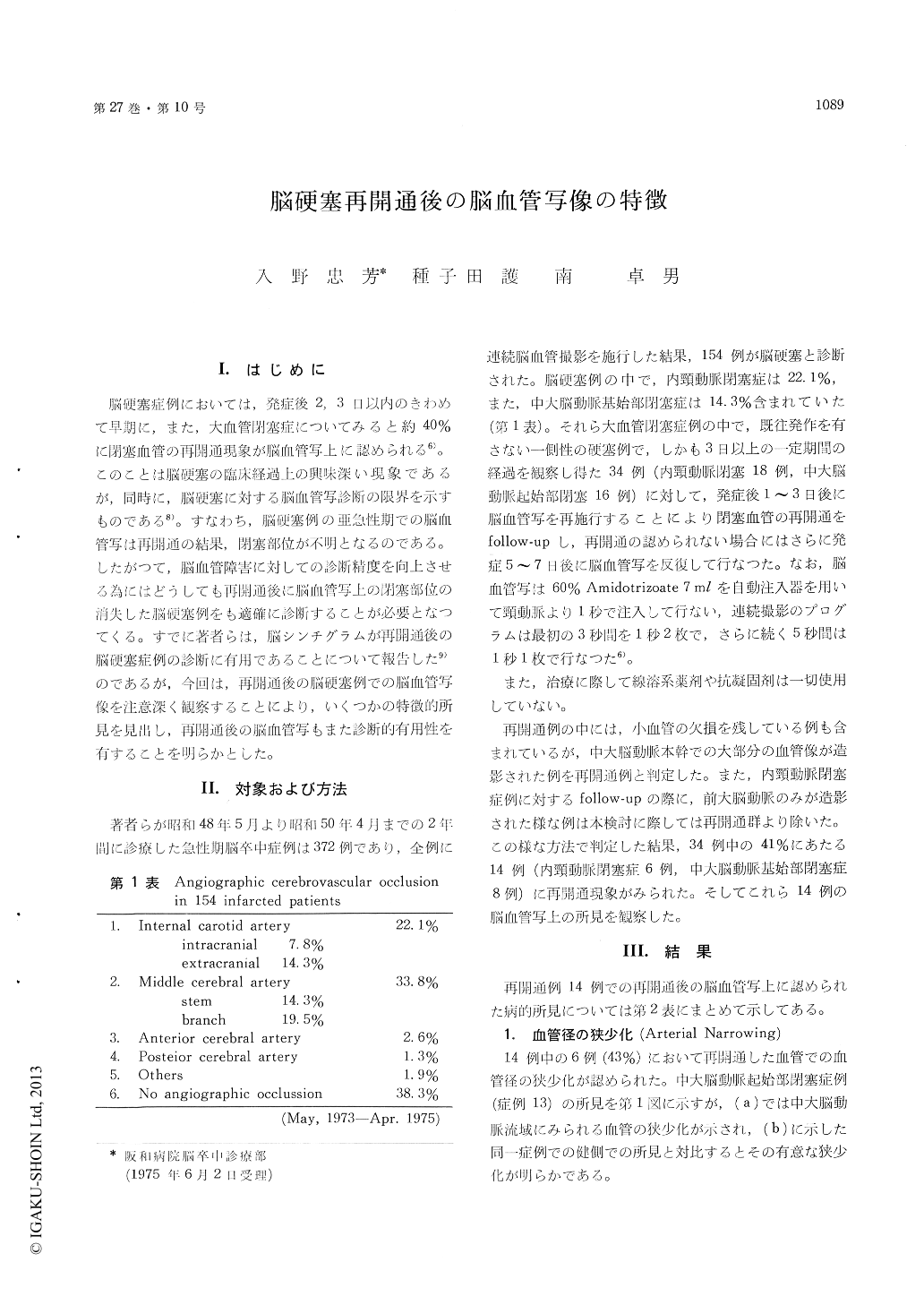
Among 154 patients diagnosed as cerebral in-farction following both physical and angiographicfindings, thirty-four patients with the internalcarotid artery or the middle cerebral arterial axisocclusion were performed consecutive cerebral angio-graphies to inspect recanalization. The follow-upangiographies were done one- to three- days intervalswithin the first week using 7 ml of Amidotrizoateserially. Consequently, six of the internal carotidarterial occlusions and eight cases with middlecerebral arterial axis occlusions were demonstratedtheir recanalization angiographically. And theangiograms of these 14 recanalized cases obtainedafter recanalization were studied carefully.
As a result, several angiographical characteristicswere revealed on the angiograms and these find-ings are summarized below. (1) Arterial narrowing;Six of the 14 recanalized cases showed narrowingof the arterial caliber, which were simulating theangiographic features of those of angiospasm. Thiswas noticeable compared with the arterial caliberon the angiograms of non-infarcted hemisphere.(2) Mass sign; Among all of the recanalized cases,eight cases showed angiographical mass sign. Inthe cases having this mass effect, three showed amid-line shift of the anterior cerebral artery withor without displacement of the middle cerebralarteries, which somewhat simulated the angiographicfindings of intracranial space occupying lesions suchas intracerebral hematoma or tumor. (3) Capillaryblush; In five cases, postrecanalized films showedmarked capillary blush in the territory of therecanalized artery. In most of cases the capillaryblush were demonstrated in the distribution of themiddle cerebral artery, while some developed thisin the area of the lenticulostriate arteries. (4)Others; Not all of the middle cerebral arterialbranches were visualized angiographically in onehalf of the recanalized cases and a few cases showedan absence of middle cerebral arterial branch likefrontoorbital branch. The residual stenosis, whichsuggested the clot lysis of the embolus, was dis-played on the arterial wall of the recanalized artery.Two cases showed significant lengthening of thecirculation time and in other two capillary phasewas rarely documented because of its short du-ration.
Summarizing the results mentioned above, thepostrecanalized angiograms had several angiographiccharacteristics such like arterial narrowing, masssign or capillary blush. Thus, the understandingthese findings may well enable the retrospectivediagnosis of recanalization even if the angiogramsat the subacute stage failed to show arterialocclusion in the infarcted patients.
Copyright © 1975, Igaku-Shoin Ltd. All rights reserved.