Japanese
English
- 有料閲覧
- Abstract 文献概要
- 1ページ目 Look Inside
I.はじめに
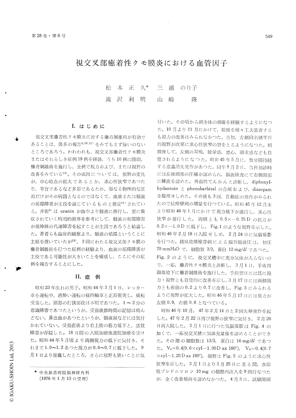
視交叉部癒符性クモ膜炎に対する癒符剥離術が有効であることは,幾多の報告5,10,11)をみてもまず疑いのないところであろう。われわれも,視交叉部癒着性クモ膜炎またはそれらしき症例19例を経験,うち10例に開頭,癒著剥離術を施行し,全例で規力および,または視野の改善をみている13)。その成因については,視野の変化が,中心暗点の拡大であるとか,求心性狭窄であつたり,半盲であるなど多彩であるため,単なる物理的な圧迫だけがその病因となるのではなくて,血液または髄液の循環障害が主役を演じているものと推定11)されている。井街4)はuraninが血中より髄液に移行し,更に吸収されていく時間的推移を参考にして,髄液の循環障害が視神経の代謝障害を起すことが主因であろうと結論した。著者らも臨床的観察より,髄液の循環ということに主眼を置いていたが13),3回にわたる視交叉部クモ膜の癒着剥離術を行つた症例の経験より,血液の循環障害が主役である可能性が大きいことを痛感し,ここにその症例を報告することにした。
A 21-year-old man struck on his head in an auto-mobile accident on March 1, 1969. Although lossof consciousness was observed for only a couple ofminutes, posttraumatic amnesia continued for sever-al hours. He had transient nasal bleeding, butliquorrhea was not noticed. About two monthsatter the accident, he complained of visual dis-turbance. Bilateral visual acuity went down fromthe previous level of 1.2 to 0.6. The pattern ofthe visual field defect advanced from left-sidedhomonymous hemianopia to concentric narrowingduring the clinical course. In Jan. 1971, visualacuity was aggravated to 0.2 on both sides. Pneumo-encephalography performed on Feb. 24 revealed thatthe air was scarcely permitted to enter the opto-chiasmatic cistern. After the adhesiotomy withmicroscorpe, the visual acuity improved upto 0.9 ontheleft side and 0.8 on the right side. Posto-perative visual fields were pointed out to be almostnormal on both sides.
On Feb. 22, 1972, the concentric limitation of thevisual fields was again noticed. Pneumoencephalo-graphy performed on March 1, apparently revealeda filling of the air in the opto-chiasmatic cistern.Intrathecal administration of steroid preparationbrought no improvement. Exploratory craniotomyas performed. There was partial fibrous adhesionamong the right internal carotid, the right anteriorcerebral artery, the chiasm and the optic nerves.After the second adhesiotomy with an operativemicroscope, preoperative pale appearance of theoptic nerves and chiasm turned into good com-plexion. Diameter of the superficial vessels on theoptic nerves and chiasm dilated macroscopically.Next day after operation a rapid recovery of thevisual acuity and fields was observed. On Apr. 10,the visual acuity showed 0.9 on the left side and0.8 on the right side, and then the test of visualfields revealed normal range. Near the middle ofDec. 1974, concentric limitation of the visual fieldswas again observed. Pneumoencephalography re-vealed air in the opto-chiasmatic cistern, however,the figure of subarachnoid space around the frontalbase and frontal pole was obscure. Therefore re-currence of the arachnoid adhesion was suspected.On Feb. 24, the third craniotomy was tried. Ad-hesion among the right internal carotid artery, theright anterior cerebral artery, the optic nerves, thetuberculum sellae and the base of the frontal lobewas found again. The complexion of the opticnerves and the chiasm improved immediately afterthe microscopical adhesiotomy Next day afteroperation the patient stated that his visual acuityand the fields recovered subjectively very well.
Following the experience of this case and thereview of literatures, the vascular factor was con-sidered to be the most important mechanism re-garding the impairment of the visual acuity andthe fields due to the adhesive arachnoiditis of theopto-chiasmatic region.

Copyright © 1976, Igaku-Shoin Ltd. All rights reserved.


