Japanese
English
- 有料閲覧
- Abstract 文献概要
- 1ページ目 Look Inside
はじめに
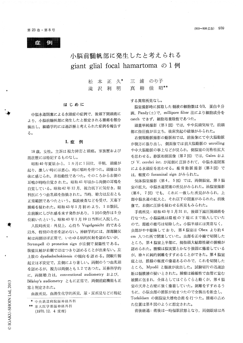
中脳水道閉塞による水頭症の症例で,後頭下開頭術により,小脳前髄帆部に発生したと推定される腫瘍を部分摘出し,組織学的には過誤腫と考えられた症例を報告する。
18-year-old girl was admitted complaining blurred vision and headache. Carotid angiography, pneumo-ventriculography and iodo-ventriculography revieled hydrocephalus due to aqueductal obstruction. Suboc-cipital craniectomy was performed, and solid tumor which was thumb tip in size was discovered in the rostral portion of the fourth ventricle. Total re-moval of the tumor was resigned because of swel-ling of the cerebellar vermis during the operation. Thereafter Torkildsen's ventriculo-cisternal shunt was performed.
In the biopsy specimen the most striking feature was that there were glial cells of various shapes and sizes. Some of them were large curious cellsand they seemed appearently like a nerve cell. However they were glial in nature and that was also verified with the Kluver-Barrera and P. T. A. H. stains. Glial fibers of the back ground showed curiously crossed coarse stream without usual fine net work. In some places proliferation of the small blood vessels was observed. The vessel walls ex-hibited no particular abnormality. Neither infarc-tion nor bleeding was observed.
We have interpreted this tumor as a hemartoma although it could not be denied completely to be a kind of giant-celled glioma.

Copyright © 1971, Igaku-Shoin Ltd. All rights reserved.


