Japanese
English
- 有料閲覧
- Abstract 文献概要
- 1ページ目 Look Inside
I.はじめに
徐々に進行する眼瞼下垂と外眼筋麻痺を主徴とするいわゆるchronic progressive external ophthalmoplegiaの大部分は,1951年Kiloh & Nevin6)が限筋組織に筋原性の変化を指摘してからは,筋ジストロフィー症の一型と考えられ,ocular myopathyと呼ばれるようになつた。しかしながら筋原性,神経原性の区別は個々の症例の場合必ずしも容易ではなく2,5),また臨床的に同様の症状を呈する他疾患との鑑別も重要である。さらに広義のocular myopathyの中には眼瞼下垂,眼球運動障害のほかに嚥下障害,四肢の筋力低下,筋萎縮などを示す症例も含まれている。すなわちLees & Liversedge8)は眼症状のほかに顔面筋,咽頭筋,喉頭筋,頸筋,上肢帯筋などが次第に侵される症例をdescending ocular myo—pathyとして報告し,Victor18)らは眠瞼下垂に嚥下障害を伴なつた例をoculopharyngeal muscular dystrophyとして報告した。また里吉ら14)は筋障寄の分布が外眼筋,舌,咽頭筋,四肢遠位部に著明な症例をoculo-phar—yngo-distal myopathyとして報告し,剖検でmyopathyであることを確認している。
これらの類似のあるいは互に共通点を示す病像が同一の原因疾患に属するものか否かは不明であるが,筋ジストロフィー症の成因の解明とも関連して種々の臨床例の積み重ねが重要である。今回我々は筋電図,筋生検などで明らかに筋原性変化を示したdescending ocular myo—pathyに視神経萎縮,網膜の機能障害,感音性難聴などの神経障害を合併した症例を経験したので報告する。
A solitary case of descending ocular myopathywas reported. This 54-year-old man was in goodhealth until about the age of 16 (1936) when droop-ing of eyelids and limitation of eye movementswere noted. These symptoms of insiduous onsetprogressed gradually, and at 20 he had a plasticoperation to raise the lids, with effect which con-tinued for only 2 to 3 years. At 44 when he hadthe same operation again, impairment of eye move-ments was almost complete. At about 50, he firstnoticed dysphonia (hoarseness and nasal voice) anddysphagia with solid food, which slowly progressed.There was neither fluctuation of the symptoms norfatigability.
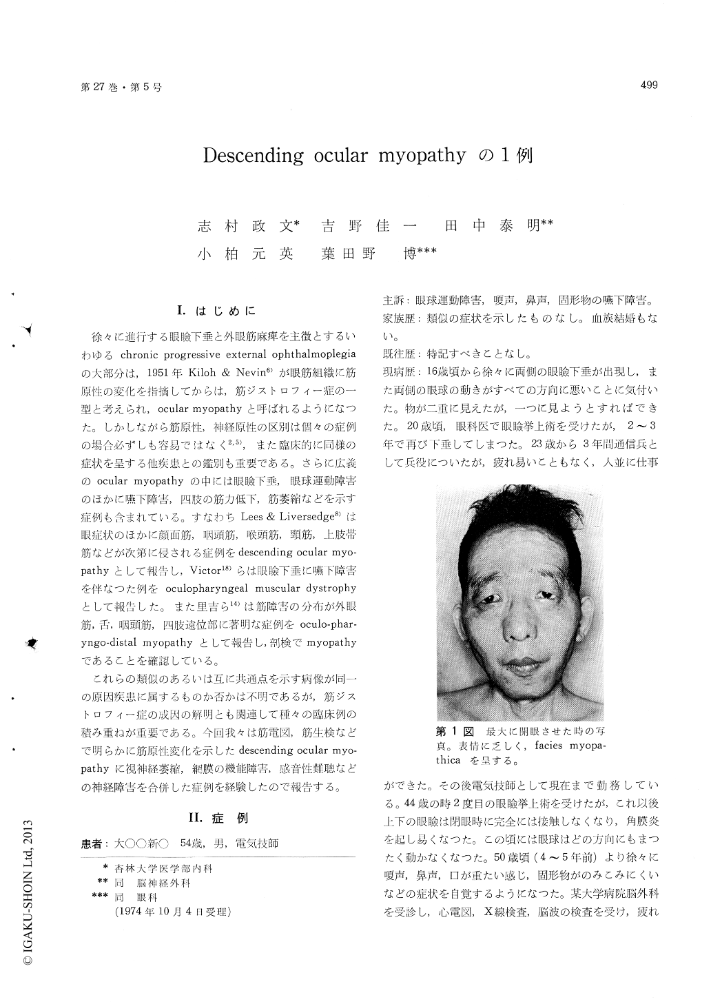
Physical examination revealed bilateral ptosis andcomplete external ophthalmoplegia. Pupillary re-action to light was normal. Facial muscles, neckmuscles especially sternocleidomastoids were allwasted and weak. Atrophy and some weaknesswere also present in the shoulder-girdle muscles,arms and to a lesser extent in the forearms andleg flexors; generally proximal muscles being af-fected more than distal muscles. Trunk muscleswere slightly weak. There was no wasting inhand muscles but moderate weakness was noted.Foot muscles were all normal. Deep tendon reflexeswere all diminished or absent. Plantar reflexeswere flexor. Sensation was normal. There wasno fasciculation. No myotonic phenomena wereobserved.
Edrophonium test was negative. Thyroid andother endocrine functions were all normal. SerumCPK was slightly elevated. There was markedcreatinuria with reduced excretion of creatinine.Skull- and chest X-ray films showed no abnormali-ties. CSF was normal. Myogenic pattern withlow voltage and short duration was obtained inEMG of orbicularis oculi and oris, masseter andsternocleidomastoid muscles. Waning was not seenon repetitive nerve stimulation. Motor nerve con-duction velocities in arms were normal. Histologyof sternocleidomastoid muscle revealed obvious my-opathic changes compatible with muscular dys-trophy.
Optic atrophy by funduscopy, impairment ofretinal function by electroretinography indicativeof some retinal degeneration and hearing loss byaudiometry were found in this case. These findingswere discussed in the light of a recent concept inthe literature concerning neurogenic symptomsconcomitant with chronic progressive externalophthalmoplegia.
Copyright © 1975, Igaku-Shoin Ltd. All rights reserved.