Japanese
English
- 有料閲覧
- Abstract 文献概要
- 1ページ目 Look Inside
I.はじめに
脳のシンチグラムはX線撮影と同じく,一平面上の投影像として表わされる。したがって,脳外の構造や組織が重なり合って投影され相互の境界が不鮮明となり,脳内病巣の形態を正確に識ることが困難な場合が少くない。具体的には,頭蓋底や後頭蓋窩の腫瘍の場合に,何れの方向の撮影を加えても副鼻腔,鼻咽頭の粘膜や前頭筋側頭筋,後頭部および頂部の筋の重複像の分離が充分に得られないことが多い。
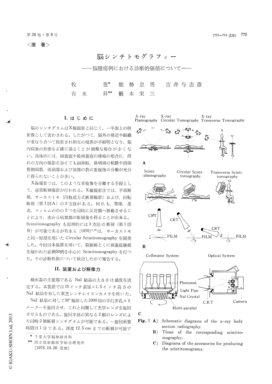
X線撮影では,このような重複像を分離する手段として,通常断層撮影が行われる。X線撮影法では,平面断層,サーカストモ(円軌道方式断層撮影)および,回転断層(第1図A)の3方法がある。何れも,管球,患者,フィルムの中の2つを同時に反対側へ移動させることにより,求める病巣部の断層像を得ることが出来る。Scintitomographyも原理的には3方法の断層(第1図B)が可能であるが有水ら(1970)1,2)は,サーカストモと同一原理を用いたCircular Scintitomographyを開発した。今回は本装置を用いて,脳腫瘍とくに頭蓋底腫瘍を疑われた症例50例を中心にScintitomographyを行った。その診断性能について検討したので報告する。
The diagnostic evaluation of the circular scinti-tomography was done 50 cases suspected brain tumors, chiefly located in the cranial floor.
We used a circular scintitomography developed by Arimizu (1970) attached Toshiba's scintillation camera, having a Nal crystal of 15 inch in diameter and 1/2 inch thick. The collimator contains 2,300 parallel round holes each inclined 30° to the face of gamma carmera crystal The rotating collimatorcan be attached and detached from the detector head as with any other collimator. Phantom studies with carmera have shown this procedure to be effective in resolving structures at various depths.
In this study, we have compared the results of scintitomogram and regular scintigrams in order to detarmine (A) whether the size and position of lesion could be defined more precisely by scintitomogra-phy. (B) whether questionable lesions and unusual anatomical variations seen on regular scintigrams could be explained by scintitomogram. (C) whether scintitomography could reveal lesions not seen in regular scintigrams.
Consequently, the scintitomography has the fol-lowing additional diagnostic aids compared with regular scintigraphy. 1) The size and site of the lesion could be more precisely discernible. 2) In the evaluation of the midline lesions, scintiomogra-phy is very useful whether the lesion is supraten-trorial or infratentorial. 3) It is very effective in the separation of the overlapping images of the extracerebral structures, which often seen in the temporal region. 4) May be helpful in the differ-enciation between the recurrence of the tumor and the radiation necrosis. 5) In a strict sense, there are no tumors which were not demonstrated in the regular scintigram but was proved to exist in thescintitomogram.
Copyright © 1974, Igaku-Shoin Ltd. All rights reserved.