Japanese
English
- 有料閲覧
- Abstract 文献概要
- 1ページ目 Look Inside
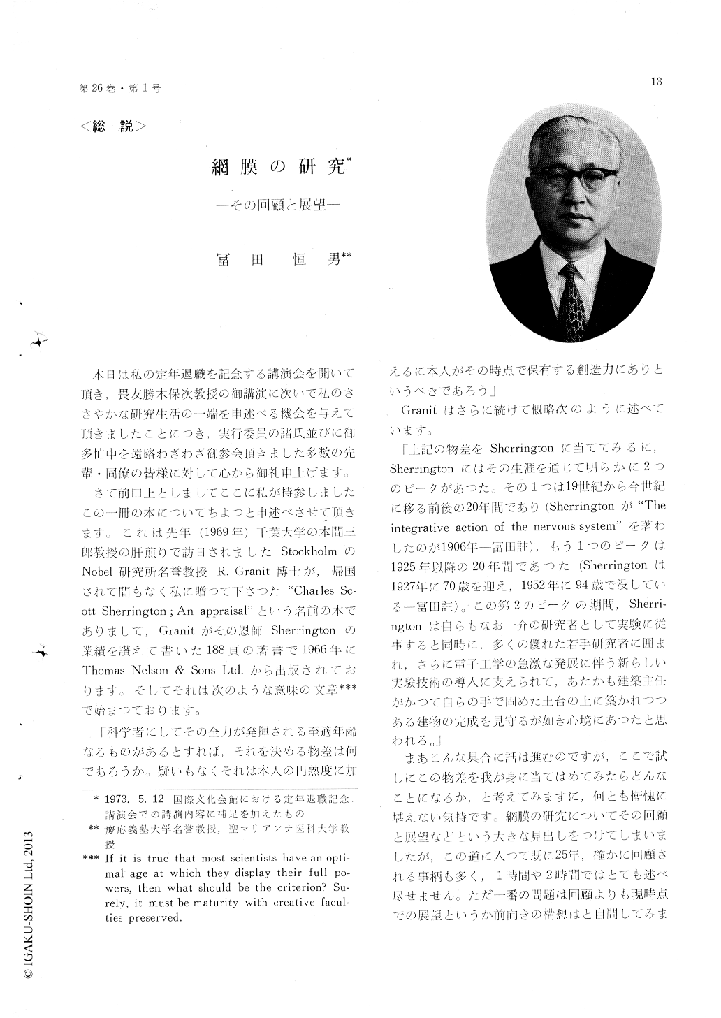
本日は私の定年退職を記念する講演会を開いて頂き,畏友勝木保次教授の御講演に次いで私のささやかな研究生活の一端を申述べる機会を与えて頂きましたことにつき,実行委員の諸氏並びに御多忙中を遠路わざわざ御参会頂きました多数の先輩・同僚の皆様に対して心から御礼申上げます。
さて前口上としましてここに私が持参しましたこの一冊の本についてちよつと申述べさせて頂きます。これは先年(1969年)千葉大学の本間三郎教授の肝煎りで訪日されましたStockholmのNobel研究所名誉教授R.Granit博士が,帰国されて間もなく私に贈つて下さつた"Charles Sc—ott Sherrington;An appraisal"という名前の本でありまして,Granitがその恩師Sherringtonの業績を讃えて書いた188頁の著書で1966年にThomas Nelson&Sons Ltd.から出版されております。そしてそれは次のような意味の文章***で始まつております。
This is an enlarged version of the lecture de-livered at a gathering in commemoration of the author's retirement from Keio University, held on May 12, 1973, at the International House, Roppongi, Tokyo.
The story begins with his attempt in 1948, three years after the end of World War II, to localizethe ERG components by means of micropipette electrodes inserted into the frog retina. The hard time is recalled, at which the experiment had to be started with substantially no equipment due to the loss of facilities by the war. The earlier ob-servations were reported in the first number of Jap. J. Physiol. (1950) and in three subsequent papers in the same journal and J. Neurophysiol., with the tentative conclusion that the main origin for the components PII and PIII is in the bipolar cell layer, whereas the component PI is of the deepest origin manifesting probably some metabolic interaction between the photoreceptors and pigment epithelial cells. The reports stimulated discussions which lasted as long as fifteen years and which brought about an important outcome that the PIII consists of two subcomponents; the distal PIII which represents the receptor potential, and the proximal PIII which originates in structures in the bipolar cell layer.
The author joyfully recalls his one year of work (1952-1953) at Professor H. K. Hartline's laboratory, Johns Hopkins. During this period, he learned the usefulness of the binocular dissection microscope and the technique of intracellular recording. In addition, he made a number of friends with whom the relation continues as good as ever.
The author's primary interest from the very beginning of his microelectrode study on the verte-brate retina was to record intracellularly the re-sponse of single photoreceptors. Such attempts, however, were never successful until 1964, the year he developed a "jolting" device which propels the tissue into the electrode at very high acceler-ations but with very small displacement. The re-sponse thus obtained from single carp cones was a sustained negative potential and definitely showed Young-Helmholtz's trichromatic type organization. He also observed that vertebrate photoreceptors work entirely differently from most other receptors. Instead of becoming depolarized by the stimulus, they are hyperpolarized. The ionic mechanism was studied with the result that the permeability for Na+ ions is high in the dark and decreased by illumination.
The recent successful application of the intra-cellular recording technique to other retinal cell types has made the analysis of their response possible. The cell identification is made after Kaneko (1970) by Procion Yellow filled electrodes. The author feels that the solution of the essential retinal mechanisms would be attained in a gener-ation. He himself works toward generalization of the observations in the retina to other nerve tissues.

Copyright © 1974, Igaku-Shoin Ltd. All rights reserved.


