Japanese
English
- 有料閲覧
- Abstract 文献概要
- 1ページ目 Look Inside
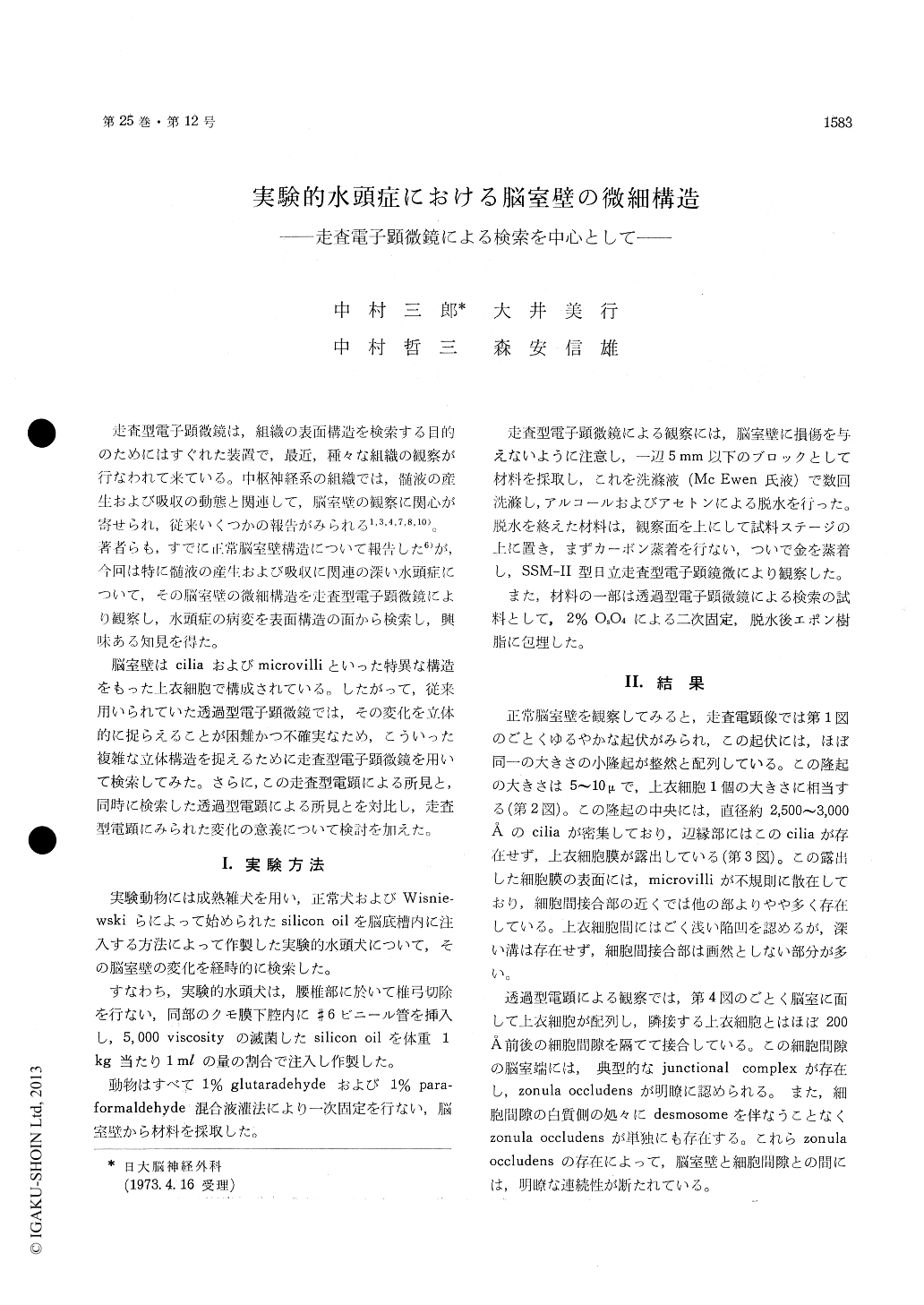
走査型電子顕微鏡は,組織の表面構造を検索する目的のためにはすぐれた装置で,最近,種々な組織の観察が行なわれて来ている。中枢神経系の組織では,髄液の産生および吸収の動態と関連して,脳室壁の観察に関心が寄せられ,従来いくつかの報告がみられる1,3,4,7,8,10)。著者らも,すでに正常脳室壁構造について報告した6)が,今回は特に髄液の産生および吸収に関連の深い水頭症について,その脳室壁の微細構造を走査型電子顕微鏡により観察し,水頭症の病変を表面構造の面から検索し,興味ある知見を得た。
脳室壁はciliaおよびmicrovilliといった特異な構造をもった上衣細胞で構成されている。したがって,従来用いられていた透過型電子顕微鏡では,その変化を立体的に捉らえることが困難かつ不確実なため,こういった複雑な立体構造を捉えるために走査型電子顕微鏡を用いて検索してみた。さらに,この走査型電顕による所見と,同時に検索した透過型電顕による所見とを対比し,走査型電顕にみられた変化の意義について検討を加えた。
Scanning electron microscopic study of hydro-cephalic ventricular wall at chronic studium, re-vealed that ependymal cell surface appeared ir-regular, cilia disappeared, interependymal groove became deeper and various openings appeared on the ventricular surface.
Ultrastructural study with a transmission elec-tron microscope showed various change in ep-endymal cells and degenerated variously. Inter-cellular junction and space of ependymal cells dissociated in many places.
In subependymal tissue appeared enlarged inter-cellular spaces and also various cavity. Sube-pendymal layer showed also extensive edema, that is, enlarged extracellular spaces, which contained clear edema fluid, and swollen myelinated nerve fiber.
Dissociated intercellular junction and enlarged intercellular spaces of ependymal layer, revealed in chronic hydrocephalus, connected ventricle to subependymal spaces continuously.
These findings suggested increased pearmeability of Ventricular wall in the hydrocephalus at chronic studium. These findings mentioned above may be one of the morphological evidences which prove that a hydrocephalus changes into arrested one.

Copyright © 1973, Igaku-Shoin Ltd. All rights reserved.


