Japanese
English
- 有料閲覧
- Abstract 文献概要
- 1ページ目 Look Inside
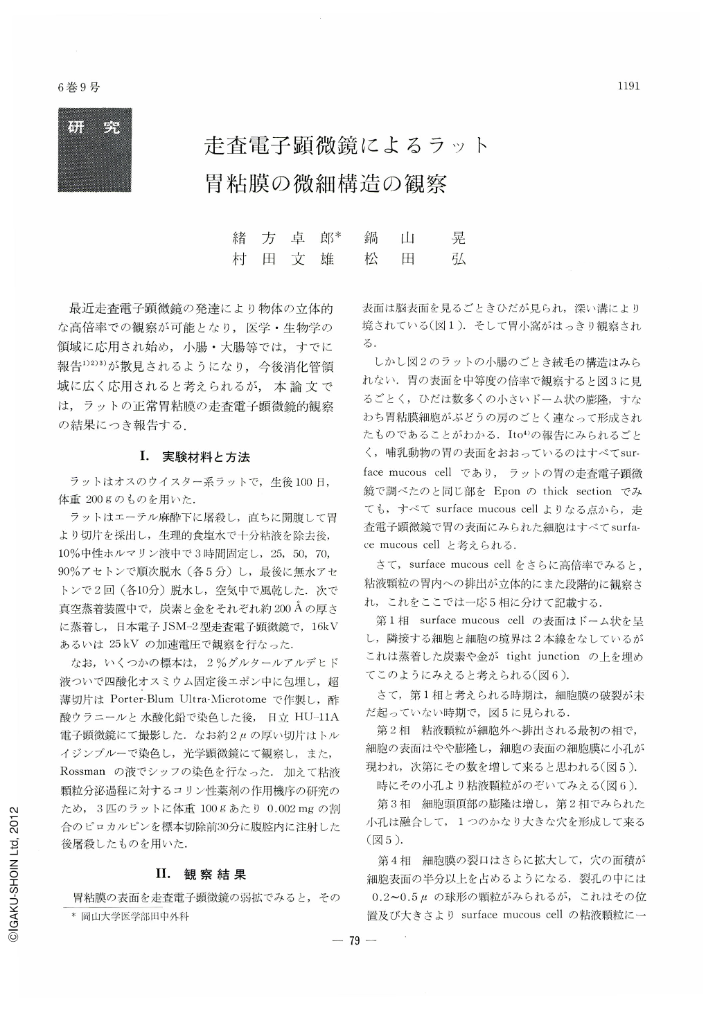
最近走査電子顕微鏡の発達により物体の立体的な高倍率での観察が可能となり,医学・生物学の領域に応用され始め,小腸・大腸等では,すでに報告1)2)3)が散見されるようになり,今後消化管領域に広く応用されると考えられるが,本論文では,ラットの正常胃粘膜の走査電子顕微鏡的観察の結果につき報告する.
The fine surface structure of the normal rat gastric mucosa was examined under the scanning electron microscope. The normal rat gastric mucosa was fixed in 10% formalin, dehydrated in acetone, dried in air, coated with carbon and gold and observed under JSM-2-type scanning electron microscope.
At the lower magnification, the mucosa formed subdivided into small bulging areas by furrows or numerous longitudinal folds or rugae, which were gastric pits. At higher magnification, the three-dimentional appearance of the secretory process on the surface mucous cell were clearly observed. The phase of the secretory process could be divided into 5 phases.
In addition, the study of the surface epithelium after pilocarpine administration reveals much more widely opened gastric pits in comparison with the control.

Copyright © 1971, Igaku-Shoin Ltd. All rights reserved.


