Japanese
English
- 有料閲覧
- Abstract 文献概要
- 1ページ目 Look Inside
はじめに
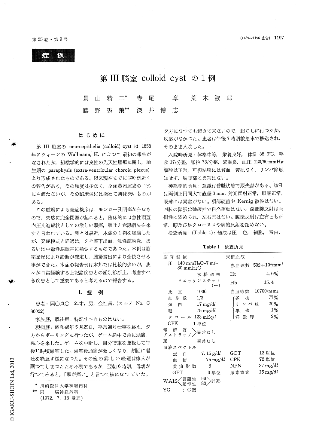
第III脳室のneuroepithelia (colloid) cystは1858年にウィーンのWallmann, H.によつて最初の報告がなされたが,組織学的には良性の先天性腫瘍に属し,胎生期のparaphysis (extra-ventricular choroid plexus)より形成されたものである。以来現在までに200例近くの報告があり,その頻度は少なく,全頭蓋内腫瘍の1%にも満たないが,その臨床像には極めて興味深いものがある。
この腫瘍による発症機序は,モンロー孔閉塞が主なもので,突然に完全閉塞が起こると,臨床的には急性頭蓋内圧亢進症状としての激しい頭痛,嘔吐と意識消失を来すと言われている。我々は最近,本症の1例を経験したが,発症模式と経過は,クモ膜下出血,急性髄膜炎,あるいは中毒性脳障害に類似するものであった。本例は脳室撮影により診断が確定し,腫瘍摘出により全快させる事ができた。本症の報告例は本邦では比較的少いが,我々が日常経験する上記諸疾患との鑑別診断上,考慮すべき疾患として重要であると考えるので報告する。
Neuroepithelial (Colloid) cyst is a benign con-genital tumor which arises from the anlage of the paraphysis, and located in the anterior superior part of the third ventricle. Clinical symptoms with attacks of headache, vomiting and altered consciousness are presumed to be due to an acute hydrocephalus produced by the block of the fora-mina of Monro.
A man, aged 21, was admitted in a state of semi-comatose following acute onset of headache, nausea and transient loss of consciousness in a single day. On admission, on May 30, 1971, he was semicoma-tose and there was urinary incontinency. Other vital signs were unremarkable. Physical examina-tion was also within normal limits. Neurological examination revealed no abnormalities of the pupils and optic fundi. There were no meningeal irri-tation signs. Limbs were flaccid and paralyzed. Deep tendon reflexes were present equally and no pathological reflexes were found. Because of non-localizing neurological signs, toxic, metabolic, or vascular disorder were considered. Lumbar punc-ture disclosed normal pressure with clear, and color-less fluid. Protein was 17 mg/dl. EEG showed paroxysmal bursts of delta waves with high voltage in all leads and the bursts were activated by photo-stimulation. After abous 24 hours of comatous state, he became slowly alert, but the amnesia persisted. Carotid angiogram revealed internal hy-drocephalus, and PEG disclosed the block the left ventricle. On 46 th hospital day, PVG was per-formed and revealed a round tumor mass (2.5 × 2.5 cm), attaching to the roof of the third ventricle. The cyst was partially removed and confirmed histologically to be the colloid cyst of the third ventricle. The patient improved withous amnesia.
Colloid cysts must be differentiated from other tumors in the third ventricle, and the differential diagnosis can not always be made before operation, unless the cysts is clealy demonstrated by ventri-culography.

Copyright © 1973, Igaku-Shoin Ltd. All rights reserved.


