Japanese
English
- 有料閲覧
- Abstract 文献概要
- 1ページ目 Look Inside
I.はじめに
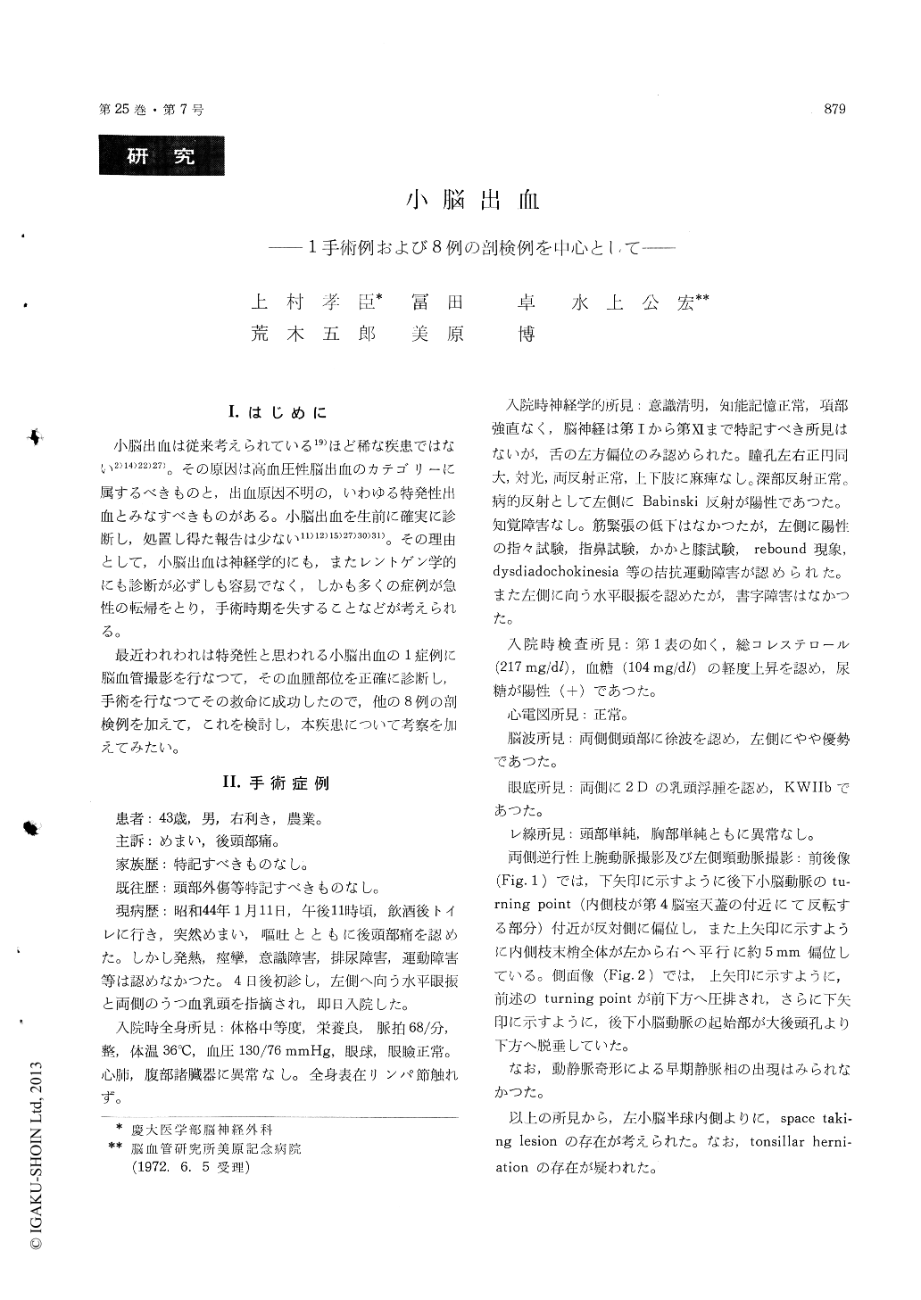
小脳出血は従来考えられている19)ほど稀な疾患ではない2)14)22)27)。その原因は高血圧性脳出血のカテゴリーに属するべきものと,出血原因不明の,いわゆる特発性出血とみなすべきものがある。小脳出血を生前に確実に診断し,処置し得た報告は少ない11)12)15)27)30)31)。その理由として,小脳出血は神経学的にも,またレントゲン学的にも診断が必ずしも容易でなく,しかも多くの症例が急性の転帰をとり,手術時期を失することなどが考えられる。
最近われわれは特発性と思われる小脳出血の1症例に脳血管撮影を行なつて,その血腫部位を正確に診断し,手術を行なつてその救命に成功したので,他の8例の剖検例を加えて,これを検討し,本疾患について考察を加えてみたい。
Cerebellar hemorrhage is classically thought to be a rare fatal disease. However, our data as well as those of others, indicate that its frequency is greater than is generally considered.
Recently we have encountered with 43 year old male who diagnosed as cerebellar hemorrhage angiographically and took satisfactory recovery by direct operative attack. Nine cases, one operated case and 8 autopsied cases, over the past 6 years were reviewed about incidence etc. with recent literatures reference.
He notes sudden onset of occipitalgia and dizzi-ness. At the time of admission neurological exa-mination revealed bilateral choked disk, nystagmus toward left, left sided dysdiadochokinesis and xanthochromia in the diagnostic spinal tap. Bi-lateral carotid angiograms showed ventricular dilatation. Bilateral retrograde brachial angio-grams revealed a space taking lesion in left cerebellar hemisphere. Preoperative pneumoven-triculography was not contributory. Suboccipital craniectomy was carried out and egg-sized intra-cerebellar hematoma was evacuated. Pathology of evacuated specimen demonstrated hemorrhage and gliosis surrounding hematoma without tumor cells or cryptic angiomatous malformations.
This case is the 4th successfully operated case in Japan. Though, all Japanese cases have chronicclinical course.
The clinical picture of all autopsied cases is not specifically characteristic comparing with those of subarachnoid hemorrhage or intracerebral hemor-rhage. The onset used to be abrupt, at times preceded by vomiting, headache and dizziness. Cerebellar signs are absent unexpectedly. The most common objective findings are initial loss of consciousness, meningeal and pyramidal signs. Blood was present in the cerebrospinal fluid in most cases. Five of patients died within the first 24 hours, 3 of them died between 8 days and 18 days.
Pathologically the highest incidence in autopsied cases was in the 5th to 6th decades. In 4 cases, location of hemorrhage was noted around dentate nucleus. Size of hemorrhage was variable from wall-nut to egg. Tonsillar herniation was noted in 4 cases. Ventricular hemorrhage was noted in5 cases. Associated lesions, though small hemor-rhage or infraction, found in the brain stem or cerebral hemisphere.
Etiologically 3 cases were attributed to hyper-tension with arteriosclerosis.
Clinically the diagnosis of cerebellar hemorrhage is quite difficult in acute stage. Most of our cases were diagnosed clinically as subarachnoid hemor-rhage. Therefore, retrograde brachial angiography is more valuable than pneumoventriculography in the point of differential diagnosis from aneurysm, arteriovenous malformation, intracerebral hema-toma etc.
We stressed the value of the retrograde brachial angiography as an useful diagnostic aid, if vessels of posterior fossa are adequately analysed by care-ful interpretation.

Copyright © 1973, Igaku-Shoin Ltd. All rights reserved.


