Japanese
English
- 有料閲覧
- Abstract 文献概要
- 1ページ目 Look Inside
I.まえがき
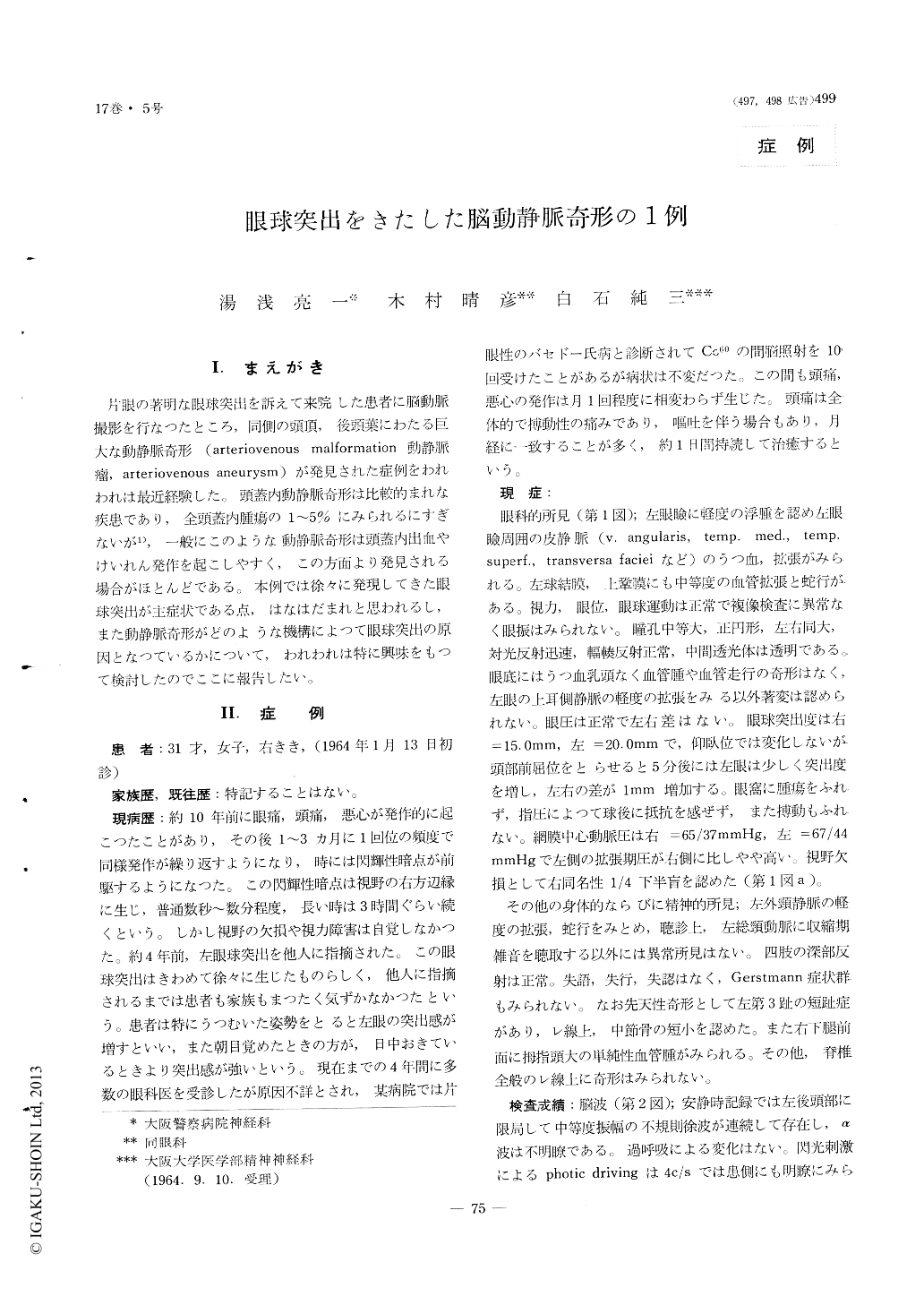
片眼の著明な眼球突出を訴えて来院した患者に脳動脈撮影を行なつたところ,同側の頭頂,後頭葉にわたる巨大な動静脈奇形(arteriovenous malformation動静脈瘤,arteriovenous aneurysm)が発見された症例をわれわれは最近経験した。頭蓋内動静脈奇形は比較的まれな疾患であり,全頭蓋内腫瘍の1〜5%にみられるにすぎないが1),一般にこのような動静脈奇形は頭蓋内出血やけいれん発作を起こしやすく,この方面より発見される場合がほとんどである。本例では徐々に発現してきた眼球突出が主症状である点,はなはだまれと思われるし,また動静脈奇形がどのような機構によつて眼球突出の原因となつているかについて,われわれは特に興味をもつて検討したのでここに報告したい。
A case of unilateral exophthalmos was presented which was resulted from the arteriovenous malfor-mation located in left parieto-occipital region.
A woman, 31-year-old, who had occasional attacks of headacke for many years, has noticed a gradual protrusion of left eye since four years. She visited to our hospital for examination on January 13,1964.
On examination, left eye was exophthalmic, mea-suring 20mm as compared to 15mm for right eye, and revealed a mederate edema of the upper and lower lids and a congestion of the conjunctiva. The exophthalmos was non-pulsative but increased slightly when she leaned forward. Left fundus revealed a normal disc with dilated veins, and a diastolic pres-sure of left central retinal artery was higher than that of right. Right inferior homonymous quadrantic anopsia was observed. The EEG showed a dysfunc-tion of the left occipital region. The Ultrasonic Blo-od-Rheograph on cerebralcirculation revealed a dec-rease of peripheral resistance, an increased blood flow in left carotid artery system and a congestive chang in left internal jugular vein.
It is considered that in this case the unilateral exophthalmos is caused by the varicosities of opht-halmic veins resulted from left internal jugular ve-nous congestion, which is caused by the increased blood flow through the arteriovenous malformation situated in left parieto-occipital region.

Copyright © 1965, Igaku-Shoin Ltd. All rights reserved.


