Japanese
English
- 有料閲覧
- Abstract 文献概要
- 1ページ目 Look Inside
I.緒言
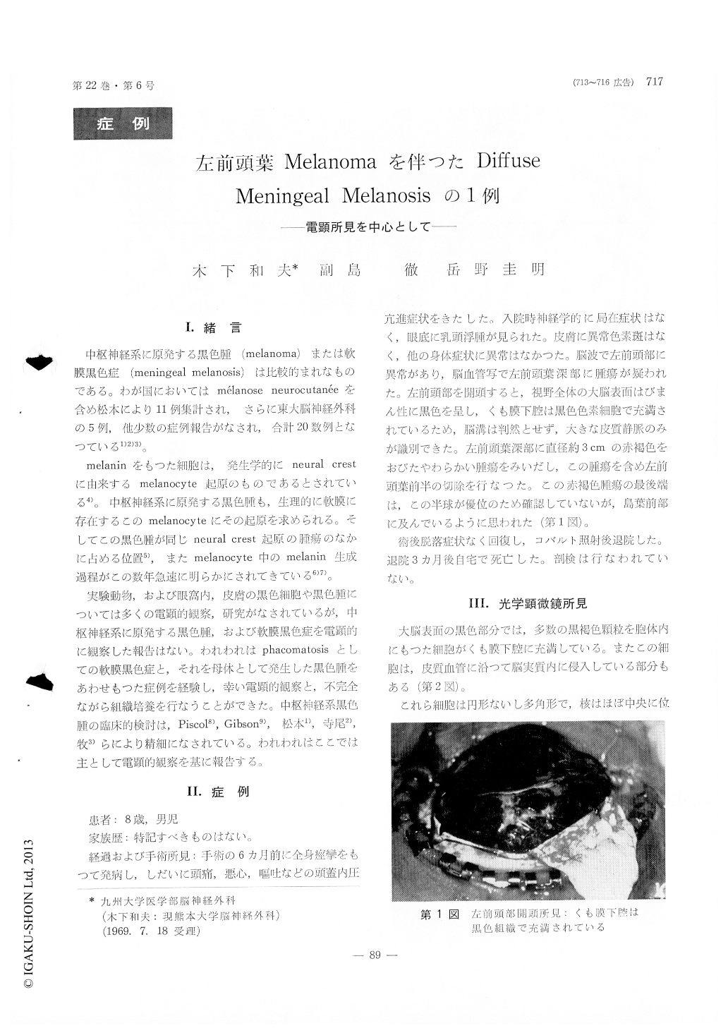
中枢神経系に原発する黒色腫(melanoma)または軟膜黒色症(meningeal melanosis)は比較的まれなものである。わが国においてはmélanose neurocutanéeを含め松本により11例集計され,さらに東大脳神経外科の5例,他少数の症例報舎がなされ,合計20数例となつている1)2)3)。
melaninをもつた細胞は,発生学的にneural crestに由来するmelanocyte起原のものであるとされている4)。中枢神経系に原発する黒色腫も,生理的に軟膜に存在するこのmelanocyteにその起原を求められる。そしてこの黒色腫が同じneural crest起原の腫瘍のなかに占める位置5),またmelanocyte中のmelanin生成過程がこの数年急速に明らかにされてきている6)7)。
The patient was an 8-year-old boy who had epi-sodes of generalized convulsive seizures, nausea, vomiting and headache of 3 months' duration. On admission he had only a papilledema still in initial stage. A left carotid angiogram demonstrated a vascular tumor in the left frontal lobe. At opera-tion the left cerebral cortex appeared diffusely black. A dark, brownish, well demarcated tumor mass, melanoma, was removed from the subcortical region in the left frontal lobe. He recovered well from surgery and was discharged without considerablesubsequent deficit after one course of Cobalt radia-tion therapy.
Light-microscopic findings :
In the region of the meningeal melanosis, the subarachnoid space was filled with round, uniform pigmented cells, which also invaded the perivascular spaces of the brain parenchyma. The melanoma cells showed a considerable pleomorphism with abun-dant mitotic figures. Many of the cells contained melanin granules in the cytoplasm in various amount. Most melanoma cells were polyhedral without pro-cesses and had a nucleus containing one or two nucleoli.
Electron-microscopic findings:
In the meningeal melanosis the cells and nuclei were uniform and contained a large amount of melanin granules. The Golgi complex was scanty. The melanoma cells showed irregular or indented nuclei, some of which had nucleoli. In few cells noted some of the epithelial character such as desmosom es and microvilli-like cytoplasmic proces-ses. In the cytoplasm there were many Golgi complexes and the melanin formation in various stages was seen in their vesicles.
Tissuse culture :
There were cells with numerous melanin granules in the cytoplasm out-growth from the tissue of the melanoma. No out-growth appeared from the tis-sue of the melanosis.
Conclusion :
From various morphological points of view it could be concluded that the diffuse meningeal mel-anosis of this case is a kind of hamartoma and the melanoma is a form of malignant change from the melanosis.
Copyright © 1970, Igaku-Shoin Ltd. All rights reserved.