Japanese
English
- 有料閲覧
- Abstract 文献概要
- 1ページ目 Look Inside
I.はじめに
原発性脳肉腫の頻度は比較的少なく,所1)によると全脳腫瘍中1%内外を占めるにすぎないが,1929年Bailey2)が(1) perithelioma,(2) perithelial sarcoma or peri—vascular sarcoma,(3) fibrosarcoma,(4) alveolarsarcomaに分類して以来,これに関する報告もかなりみられるようになつた。なかでも細網内皮系肉腫は脳で部は比較的まれなものとされており,従来primary mesen—chymal sarcoma, stem cell sarcoma, perithelial sarco—ma, microglioma, reticulum cell sarcomaなど種々の名称でよばれており,いずれもその細胞発生に対する考えかたに由来するもので,この点に関しても興味のあることである。
われわれは最近脳内に原発した細網内皮系肉腫3例,うち1例はいわゆるmicroglioma,ほか2例はreticulumcell sarcomaと考えられる症例を剖検により経験し,これらをあわせ検討する機会を得たので主としてその病理学的所見を述べ,あわせて若干の文献的考察を加え報告する。
In the primary sarcomas of the brain, attention was paid to the neoplasms of the reticuloendothelial system in relation to the origin of this neoplasm. Three autopsy cases of tumor of this type were reported in the present study.
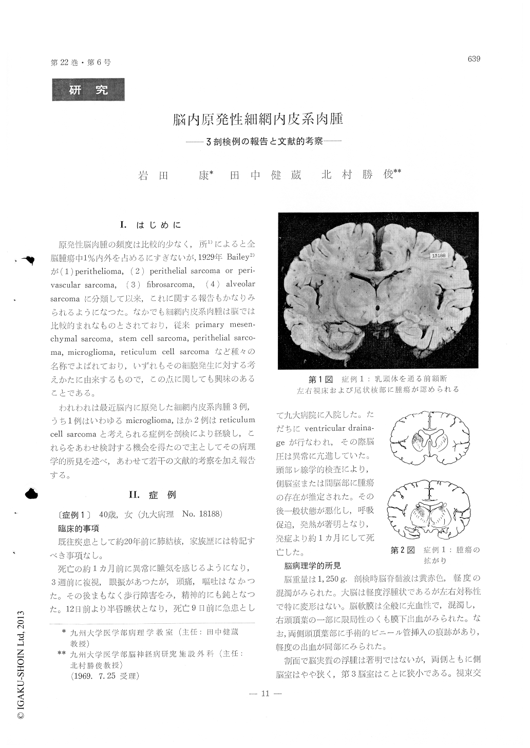
The first case, a 40-year-old woman, took very short downhill course. The tumor tissues occupied both sides of the basal ganglia. In the second case, a 63-year-old man, many tumor masses distributed in the basal ganglia,the cerebral hemispheres and the base of the fourth ventricle. In a 56-year-old man, the third case, a tumor mass was surgically removed from the right parietal lobe about five months prior to death. Post-mortem examination revealed an-other tumor mass at the left fronto-parietal lobe, without a recurrence at the site of the removal.
Histological examination revealed that the neo-plasm of the first case was composed of small round cells with scanty cytoplasm and hyperchromatic nuclei, showing prominent mitotic figures and peri-vascular arrangement with numerous reticulin fiber formation. Microglial stainning method (Weil-Davenport, Penfield) proved microglial elements in many of the tumor cells. On the other hand, swollen and phagocytic microglial cells were also found in the tumor tissues. Histological findings of the other two cases showed the same pattern as that of the reticulum cell sarcoma. The tumor cells were chiefly polygonal and pleomorphic. Gomori's silver impregnation disclosed intertwined arrange-ment of reticulin fibers to the tumor cells and peri-vascular proliferation with mesh of reticulin network. Silver carbonate stainning failed to demonstrate metallophilic mieroglia in these tumor cells of both cases.
The first case was diagnosed as microglioma and the second and third cases as reticulum cell sarcoma. All these neoplasms seemed to be originated in the reticuloendothelial component of the perivascular space in the brain.

Copyright © 1970, Igaku-Shoin Ltd. All rights reserved.


