Japanese
English
- 有料閲覧
- Abstract 文献概要
- 1ページ目 Look Inside
I.はじめに
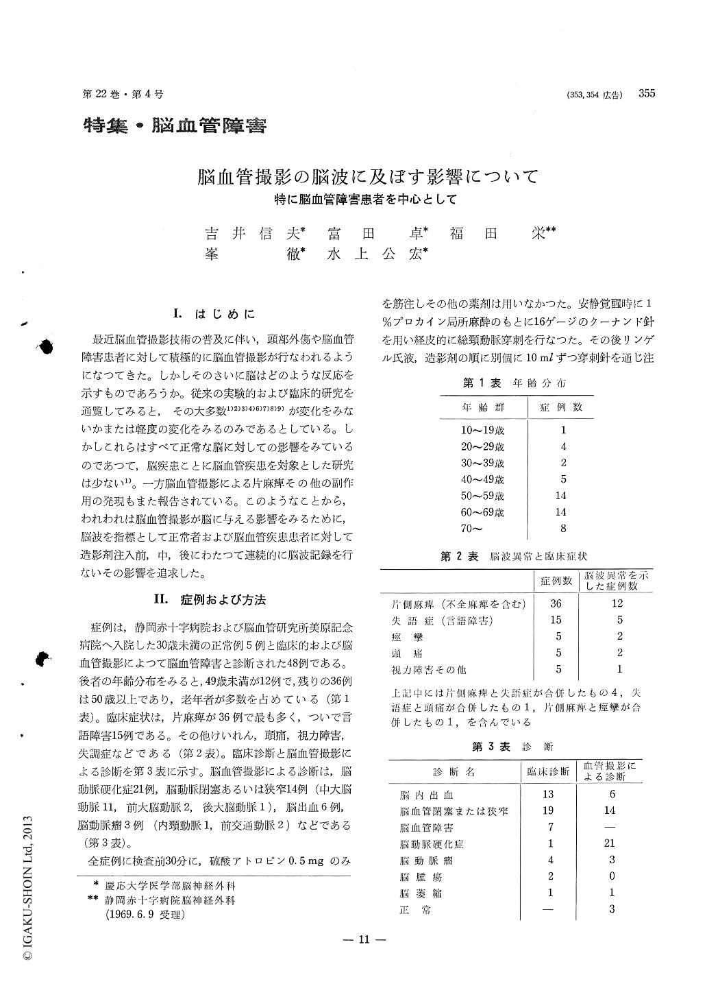
最近脳血管撮影技術の普及に伴い,頭部外傷や脳血管障害患者に対して積極的に脳血管撮影が行なわれるようになってきた。しかしそのさいに脳はどのような反応を示すものであろうか。従来の実験的および臨床的研究を通覧してみると,その大多数1)2)3)4)6)7)8)9)が変化をみないかまたは軽度の変化をみるのみであるとしている。しかしこれらはすべて正常な脳に対しての影響をみているのであつて,脳疾患ことに脳血管疾患を対象とした研究は少ない1)。一方脳血管撮影による片麻痺その他の副作用の発現もまた報告されている。このようなことから,われわれは脳血管撮影が脳に与える影響をみるために,脳波を指標として正常者および脳血管疾患患者に対して造影剤注入前,中,後にわたって連続的に脳波記録を行ないその影響を追求した。
Electroencephalographic study of patients sub-jected to carotid angiography using Urographin & Conrey failed to show siginificant alteration of the tracing as result of contrast medium.
1) Five normal adult cases revealed no changes between pre and post-angiographic EEG tracing.
2) Four CVA cases gave EEG changes after the injection of Ringer's solution into the carotid artery.
3) 33% of CVA cases revealed EEG changes during and after the angiography with contrast medium.
4) Details of EEG finding were as follows ; i) in-creased focal abnormality in 13 cases including 5 cases of new focal formation. ii) Background activity changes such as decreased frequency & increased or decreased amplitude were seen in 4 cases, iii) One case had bilateral diffuse burst.
Cause of these EEG changes were considered as cerebral metabolic alteration due to stimulus & con-sistency of contrast medium & sudden pressure change during angiography.

Copyright © 1970, Igaku-Shoin Ltd. All rights reserved.


