Japanese
English
- 有料閲覧
- Abstract 文献概要
- 1ページ目 Look Inside
緒言
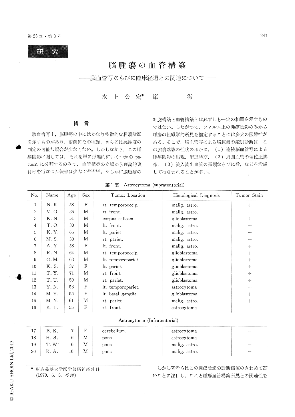
脳血管写上,脳腫瘍の中にはかなり特徴的な腫瘍陰影を示すものがあり,術前にその種類,さらには悪性度の判定の可能な場合が少なくない。しかしながら,この腫瘍陰影に関しては,それを単に形態的にいくつかのpa—tternに分類するのみで,血管構築の立場から理論的裏付けを行なった報告は少ない5)11)13)。たしかに脳腫瘍の細胞構築と血管構築とは必ずしも一定の相関を示すものではない。したがって,フィルム上の腫瘍陰影のみから腫瘍の組織学的所見を推定することには多大の困難性がある。そこで,脳血管写による脳腫瘍の鑑別診断は,この腫瘍陰影の性状のほかに,(1)連続脳血管写による腫瘍陰影の出現,消退時期,(2)周囲血管の偏位圧排像,(3)流入流出血管の種類ならびに数,などを考慮して行なわれることが多い。
Angioarchitecture of twenty astrocytomas (Kernohan's classification grade 1-grade 4) hasbeen studied with the use of Hasegawa-Ravens method and microangiography. These findings, in turn, have been correlated with the serial angiograms and biological malignancy.
1) Angioarchitecture of the astrocytoma is characterized by the alternation of normal capil-lary networks, increase in vascular number, tor-tuosity and dilatation of vessel caliver. And it would seem that some correlation exists between these vascular pattern gradations and the degree of cytological malignancy. In glioblastoma multi-forme, the vascular pattern became chaotic and numerous large vessels with the diameter of more than handreds micra were observed. In some cases of glioblastoma multiforme anastomotic channels and glomeruloid formations were visible.
2) Angioarchitecture of the astrocytoma differs depending on their origin and manner of the growth. The astrocytoma within or infiltrating into the cerebral cortex where capillary networks are more numerous shows more increased vas-cularity than that within white matter or brain stem.
3) In malignant astrocytoma, angioarchitecture varies from one part of the tumor to another. There is a gradual transition from normal capil-laries at the periphery of the tumor to the dila-tation and tortuosity of the inner part of it. It would, therefore, appear that the tumor staining in cerebral angiograms does not necessarily indi-cate true size of the tumor. This observation is an important fact for the successful resection and radiation therapy of the tumor.
4) The astrocytoma was classified into three groups by the grade of angioarchitecture and angiographic picture as follows, i) astrocytoma, ii) malignant astrocytoma and iii) glioblastoma multiforme. It can be said that the rapider the tumor growth, the more dilated and tortuous the tumor vessels. And clinical malignancy depends upon the degrees of increased tumor vascularity, dilatation of preexisting vessel and arteriovenous shunt in cerebral angiogram. We would like to stress that these findings are very important in the diagnosis and treatment of the astrocytoma.
5) It is difficult to make a diagnosis of the astrocytom from angiographic pictures alone. But angioarchitecture of the brain tumor shows essen-tially very different patterns according to their species in our experience. The study of angio-architecture of the brain tumor may aid, there-fore, in better recognition of the tumor and assist in the differential diagnosis of them in cerebral angiograms.

Copyright © 1971, Igaku-Shoin Ltd. All rights reserved.


