Japanese
English
- 有料閲覧
- Abstract 文献概要
- 1ページ目 Look Inside
I.緒言
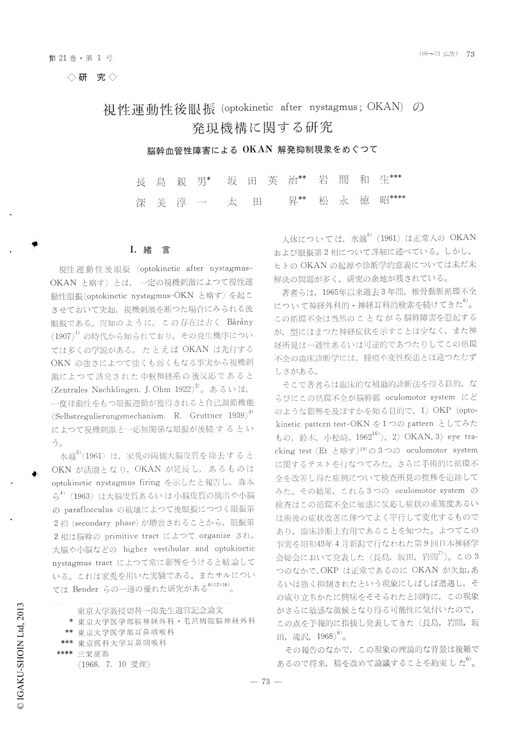
視性運動性後眼振(optokinetic after nystagmus—OKANと略す)とは,一定の視機刺激によって視性連動性眼振(optokinetic nystagmus-OKNと略す)を起こさせておいて突如,視機刺激を断つた場合にみられる後眼振である。周知のように,この存在は古くBarany(1907)1)の時代か知られており,その発生機序については.多くの学説がある。たとえばOKANは先行するOKNの強さによつて強くも弱くもなる事実から視機刺激によつて誘発された中枢神経系の後反応であると(Zentrales Nachklingen. J. Ohm 1922)2)。あるいは,一度律動性をもつ眼振運動が獲得されると自己調節機能(Selbstregulierungsmechanism. R. Gruttner 1939)3)によつて視機刺激と一応無関係な眼振が後続するという。
水越5)(1961)は,家兎の両側大脳皮質を除去するとOKNが活溌となり,OKANが延長し,あるものはoptokinetic nystaglnus firingを示したと報告し,森本ら4)(1963)は大脳皮質亜あるいは小脳皮質の摘出や小脳のparaflocculusの破壊によって後眼振につづく眼振第2相(secondary phase)が増強されることから,眼振第2相は脳幹のprimitive tractによってorganizeされ,大脳や小脳などの higher vestibular and optokinetic nystagmus tractによって常に影響をうけると結論している。これは家兎を用いた実験である。またサルについてはBenderらの一連の優れた研究がある8)12)18)。
Defective optokinetic after nystagmus (OKAN) had been frequently observed by the authors in cases of vertebro-basilar insufficiency. This defective OKAN is called even when the preceding optokinetic nys-tagmus (OKN) is normally active, the OKAN alone, is abolished or abated as shown in Fig. 1 and 2. In order to clearify the underlying neuronal mechanism of this defective OKAN in man, authors attempted to have simultaneous recording of EEG and ENGin normal subjects upon testing OKN and OKAN utilizing Ohm type apparatus (Fig. 3). OKAN was observed on extinguish the light. The results as follows.
1) OKAN was observed following total darkness when EEG had returned to alpha wave activity after short interval of desynchronized pattern (Fig. 5). This indicates the cortical cells are not engaged in any specific task to form the OKAN. 2) On changing to the secondary phase of OKAN, at the point where inversion of the after nystagmus had taken place, the EEG does not show any change of it's alpha wave pattern. This represents the cortical cells are not engaged in any task to form the secondary phase of after nystagmus. 3) In a subject with excellent visual acuity to follow moving object showed two different discreete patterns in EEG depending upon quick and slow phase of OKN-arousal pattern in slow phase and alpha rhythm in quick phase (Fig. 4). This indicates the cortical cells are not engaged to any task to from the quick phase of OKN. 4) From the facts mentioned above, the authors concluded that OKAN, secondary phase of OKAN, quick phase of OKN are organized from suhcortical mechanism, probably from the oculomotor system in the brain stern.

Copyright © 1969, Igaku-Shoin Ltd. All rights reserved.


