Japanese
English
- 有料閲覧
- Abstract 文献概要
- 1ページ目 Look Inside
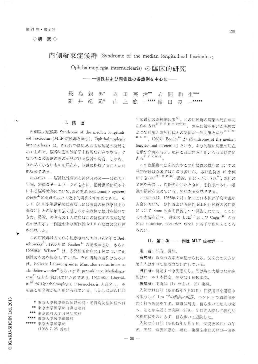
I.緒言
内側縦束症候群Syndrome of the median longitudi—nal fasciculus (MLF症候群と略す),Ophthalmoplegia internuclearisは,きわめて特長ある眼球運動の所見を示すもので,脳幹障害の診断学上特異な存在である。すなわちこの眼球運動の所見だけで脳幹の病変,しかも,きわめて小さいものの局在を,的確に指摘することが可能なのである。
われわれ—脳神経外科医と神経耳科医—は過去3年間,密接なチームワークのもとに,椎骨動脈循環不全による脳幹障害について,眼運動系(oculomotor system)の観察1)に重点をおいて臨床的研究をすすめてきた。そして《この眼運動系の観察なしには脳幹の神経学はあり得ない》との印象を強く感じながら症例の検討を続けてきた。最近,著者らの1人長島はこの特徴ある眼球運動の所見を示す一側性および両側性MLF症候群の各症例を発見した。
This paper consists of case presentation of syn-drome of the medial longitudinan fasciculus (MLF syndrome) and analytical review of reported autopsy cases of this syndrome drawn from the literature.
Report of cases
Case 1. Sudden onset of vertigo, vomiting, diplo-pia, unilateral MLF syndrome with post-traumatic onset.
A 52-year-old male was admitted to Moro-General Hospital because of sudden onset of vertigo, vomiting and diplopia. He had had injury to the forhead by automobile accident without loss of consciousness 10 days prior to the onset. The onset of the present symptoms had been sudden, while he was sitting on a chair after dinner with severe attack of vertigo, vomiting, diplopia which gradually disappeared on the next morning. Recurrence of the same attack occured in the next evening after drinking 1 2 glass of beer. He was admitted 2 days after initial attack. Examination disclosed complete absence of adduction of the left eye on attempted conjugate gaze but nor-mal adduction on convergence. The right eye display-ed monocular horizontal nystagmus with full abdu-ction. Vertical nystagmus directed upward and down-ward was noted. This was marked seen through the Frenzel glass or upon testing positioning nystagmus of Dix-Hallpike and Stenger. Electronystagmogram with open eyes looking forward showed alternatingburst of horizontal nystagmus with frequency of 3-4 c. p. s. and duration varying 0. 8 sec. to 4. 0 sec. in one direction. The direction of nystagmus was al-ternating quite irregularly. We denoted this phenom-ena as irregular alternating burst of horizontal nystagmus and attributed this phenomena to the lesion in the paramedian reticular formation in the pons. There was no other disturbance of ocular movements, no ptosis, no pupillary abnormality. There was no obvious paresis of the face, arms or legs ; the plantar reflexes were normal, and no other abnormal neurological signs were elicited. The cereb-ral angiography through the retrograde brachial artery showed atheromatous irregular narrowing both in the internal carotid and vertebral artery i. e. at the carotid syphon and the area where the poste-rior inferior cerebellar artery originates. Transient central facial nerve paresis was followed post angio-graphy on the same side with MLF lesion, which had completely recovered for 2 weeks. By the 4 weeks after the onset. ocular abnormality had com-pletely disappeared. Twelve-months followup reveal-ed no further neurological abnormality.
Case 2 Bilateral MLF syndrome, conjugate gaze palsy to upward and downward, loss of convergence, in a patient after normal delivery.
A 23-year-old female, came to our clinic because of diplopia, blurring of vision and frontal headache of 6 days' duration. These symptoms begun 5 days after normal delivery on looking far distance and were accompanied transient numbness in her left fingers and bilateral ptosis. No vertigo or dizziness.
Examination disclosed complete absence of adduc-tion of the adducting eye on attempted lateral gaze with monocular horizontal nystagmus of abducting eyes on attempted conjugate lateral gaze to the left and right. There were complete paralysis of up-ward and downward gaze and loss of convergence on command. Mild ptosis persisted. On attempted downward gaze there was vertical nystagmus directed downward. The pupils were equal in size and reacted prompt to light and did not react to near vision. Fundoscopic examination showed no abnormality. No visual field defect. No deviation, no Homer's sign. The neurological status was otherwise nor-mal. The Tensilon test to rule out myasthenia gravis gave a negative reaction. One week after the onset, vertical movements of eyes begun to improve with occasional convergence nystagmus at the time. No retraction nystagmus, no convergence spasm accompanied. On three weeks after the onset both eyes adducted well and monocular horizontal nystagmus was not elicited. By the one month after the onset all of ocular findings disappeared. The patient has now been followed for 9 months. There have been no attack of diplopia, no neurological deficit.
Analytical review of reported autopsy cases
Ten reported autopsy cases of MLF syndrome including of Spiller (1924), Sahlgren, et al. (1950) Congan, et al. (1950) Case Record of Massachusetts General Hospital (1953) Gosztonyi (1961) Ross (1966) Christoff, at al. (1966) Kupfer, et al. (1966) were tabulated in Table 1, 2, 3. Evidence from these clinicopathological cases suggests as follows ; (1) The essential features of the syndrome are paresis of adduction of ipsilateral eye and monocular horizon-tal nystagmus in the abducting eye on conjugate lateral gaze, convergence preserved. (2) Other types of ocular dysfunction such as skew deviation, paresis of upward or downward gaze, ptosis are due to injury of surrounding structures and not due to the MLF lesion itself. (3) There noted no signifi-cant difference in ocular manifestation depending upon level of lesion in the MLF.

Copyright © 1969, Igaku-Shoin Ltd. All rights reserved.


